Review
. 2020 Dec;17(12):659-678.
doi: 10.1038/s41585-020-00382-9. Epub 2020 Oct 13.

Sarcomatoid renal cell carcinoma: biology, natural history and management

Affiliations
Review

Sarcomatoid renal cell carcinoma: biology, natural history and management

Kyle A Blum et al. Nat Rev Urol. 2020 Dec.

Abstract

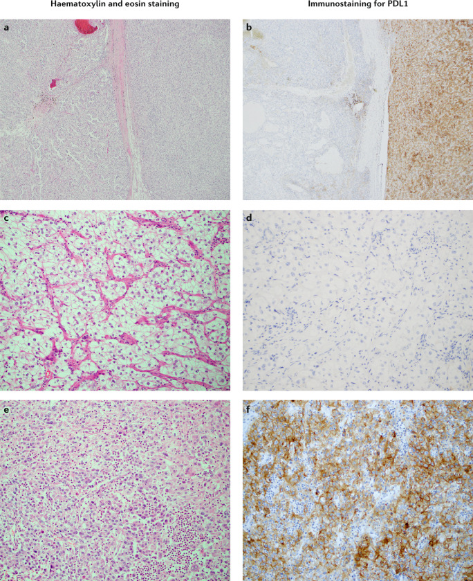
Sarcomatoid dedifferentiation is an uncommon feature that can occur in most histological subtypes of renal cell carcinomas (RCCs) and carries a decidedly poor prognosis. Historically, conventional treatments for sarcomatoid RCCs (sRCCs) have shown little efficacy, and median survival is commonly 6-13 months. Despite being first described in 1968, the mechanisms driving sarcomatoid dedifferentiation remain poorly understood, and information and treatment options available to physicians and patients are limited. When diagnosed at an early stage, surgical intervention remains the treatment of choice. However, preoperative identification through routine imaging or biopsy is unreliable and most patients present with advanced disease and systemic symptoms. For these patients, the role of cytoreductive nephrectomy is disputed. The expansion of immunotherapies approved for RCCs has generated a search for biomarkers that might be indicative of treatment response in sRCCs, although a proven effective systemic agent remains elusive. PDL1 expression is increased in sarcomatoid dedifferentiated renal tumours, which suggests that patients with sRCCs could benefit from PD1 and/or PDL1 immune checkpoint blockade therapy. Treatment outcomes for sarcomatoid tumours have remained relatively consistent compared with other RCCs, but further investigation of the tumour-immune cell microenvironment might yield insights into further therapeutic possibilities.


Conflict of interest statement

J.A.K. is a member of consulting or advisory boards for, and has received honoraria from, Merck, EMD Serono, Genentech, Novartis and Pfizer. J.A.K. has received institutional research funding from Merck, Genentech and Mirati. R.J.M. has received grants and paid consultancy from Eisai, Exelixis, Genentech, Roche, Novartis, Merck and Pfizer, paid consultancy from Astra Zeneca and grants from Bristol Myers Squibb. All other authors declare no competing interests.

Figures

Fig. 1
Fig. 1. Gross sections of an sRCC after radical nephrectomy.
Sarcomatoid renal cell carcinoma (sRCC) components are often large and can appear as dense grey or white areas within the tumour architecture and typically reveal a firm and fleshy cut surface when dissected. Arrow shows fleshy area that corresponds to sarcomatoid transformation and asterisk marks yellow and friable area of the tumour that corresponds to lower grade clear cell renal cell carcinoma.
Fig. 2
Fig. 2. Histopathology of sRCC.
Part a (×40 magnification) shows a representative area of a sarcomatoid renal cell carcinoma (sRCC) with a well-differentiated clear cell carcinoma (ccRCC) component (asterisk) and a dedifferentiated sarcomatoid component (arrow). Magnified areas of the ccRCC (part b, ×200 magnification) and dedifferentiated sarcomatoid components (part c, ×200 magnification) are also shown. The ccRCC comprises cells with optically clear cytoplasm organized in alveolar/acinar architectural patterns, whereas the sarcomatoid component exhibits spindled cells associated with a dense lymphocytic infiltrate. Immunohistochemistry for carbonic anhydrase IX (a HIF-1α target gene that shows diffuse membranous localization in ccRCCs, which have increased HIF-1α signalling secondary to alterations of VHL) shows an area of transformation (part d, ×40 magnification), with strong cell membrane-localized expression in the well-differentiated ccRCC areas (asterisk) and gradual loss of expression in the more poorly differentiated sarcomatoid areas (arrow).
Fig. 3
Fig. 3. Signalling pathways involved in EMT reported in sRCC.
A sarcomatoid renal cell carcinoma (sRCC) is composed of two separate cell types, the sarcomatoid (mesenchymal) component, and the RCC (epithelial) component. The mechanism by which sarcomatoid dedifferentiation arises within RCC is not clearly understood; however, there is evidence that the sarcomatoid component may originate from a common cell-of-origin, resulting in cells that lose their epithelial characteristics and gain mesenchymal characteristics through a process called epithelial–mesenchymal transition (EMT). EMT can occur via multiple pathways including TNF, TGFβ, Wnt, MAPK and PI3K/AKT signalling to regulate expression of Snail, Zeb and Twist. Activation of these transcription factors results in the downregulation of epithelial markers (E-cadherin) and upregulation of mesenchymal markers (N-cadherin). E-cadherin is a cell membrane protein that is important in cell–cell adhesion; however, during EMT these intercellular tight junctions (E-cadherin) break down and transform the cell into a more mesenchymal phenotype that increases the likelihood of tumour cell metastasis.
Fig. 4
Fig. 4. PDL1 expression in sRCC.
Representative haematoxylin-and-eosin-stained images (left) of a 9p24.1-amplified clear cell renal cell carcinoma (ccRCC) with sarcomatoid transformation are depicted alongside corresponding immunostaining for PDL1 (right). Staining for PDL1 is absent in areas with a clear cell component and shows constitutive expression in areas with a sarcomatoid component, which is higher grade than the clear cell component. Constitutive expression of PDL1 in higher grade sarcomatoid components implies an underlying molecular event such as an amplification in JAK2, PDL1 and PDL2 at the 9p24.1 locus. Patients with this expression pattern have the potential for an enhanced response to immune checkpoint inhibitors owing to the increased expression of PDL1. Parts a and b show adjacent clear cell and sarcomatoid areas (×40 magnification). Parts c and d show a representative area with clear cell morphology (×200 magnification). Parts e and f show a representative area with sarcomatoid morphology (×200 magnification). Reprinted from ref., Springer Nature Limited.

References

    1. Siegel RL, Miller KD, Jemal A. Cancer statistics, 2019. CA Cancer J. Clin. 2019;69:7–34. - PubMed
    1. Capitanio U, et al. Epidemiology of renal cell carcinoma. Eur. Urol. 2019;75:74–84. - PMC - PubMed
    1. Bray F, et al. Global cancer statistics 2018: GLOBOCAN estimates of incidence and mortality worldwide for 36 cancers in 185 countries. CA Cancer J. Clin. 2018;68:394–424. - PubMed
    1. Motzer RJ, et al. Kidney cancer, version 2.2017, NCCN clinical practice guidelines in oncology. J. Natl Compr. Canc. Netw. 2017;15:804–834. - PubMed
    1. Delahunt B. Sarcomatoid renal carcinoma: the final common dedifferentiation pathway of renal epithelial malignancies. Pathology. 1999;31:185–190. - PubMed

Publication types

MeSH terms