

Contralateral pulmonary resection using selective bronchial blockade in postpneumonectomy patients
- PMID: 33052015
- PMCID: PMC7705631
- DOI: 10.1111/1759-7714.13696
Contralateral pulmonary resection using selective bronchial blockade in postpneumonectomy patients
Abstract
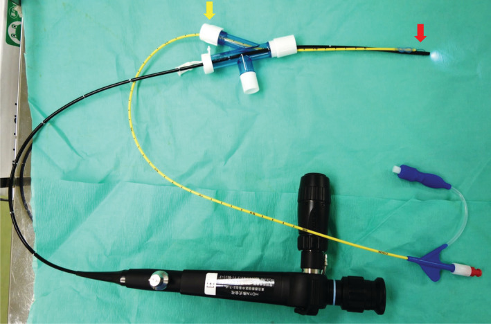
Background: Pulmonary resection is occasionally performed in postpneumonectomy patients with contralateral lung lesions, such as metachronous or metastatic lung cancer. Careful intraoperative respiratory management is essential in such patients. This study evaluated the respiratory management of postpneumonectomy patients who underwent contralateral pulmonary resection with selective bronchial blockade of the lobe or segment to be resected.
Methods: We retrospectively analyzed the surgical findings and safety of surgery in six patients who underwent contralateral pulmonary resection with selective bronchial blockade after pneumonectomy for non-small cell lung cancer (NSCLC).
Results: The percutaneous oxygen saturation did not decrease in any of the patients during bronchial blockade under high oxygen concentration. The median blockade time was 57.5 minutes. The operative field was tolerable secured under conditions of partial lung collapse, and partial pulmonary resection was performed as planned. Postoperatively, one patient developed acute respiratory distress syndrome due to acute exacerbation of interstitial pneumonia; however, no patients died within one month postoperatively. Two patients underwent pulmonary resection in order to obtain adequate tissue specimens to evaluate the biomarkers of multiple lung metastases. On histopathology, one patient tested positive for anaplastic lymphoma kinase (ALK) and was subsequently administered an ALK inhibitor, which prolonged survival.
Conclusions: In all patients, intraoperative respiratory condition under partial lung collapse remained stable, and all partial pulmonary resections were safely performed. However, surgical indications should be carefully reviewed preoperatively in patients with interstitial pneumonia.
Key points: SIGNIFICANT FINDINGS OF THE STUDY: Contralateral partial pulmonary resection was performed using selective bronchial blockade in postpneumonectomy patients. Percutaneous oxygen saturation did not decrease during the bronchial blockade under high oxygen concentration, and the operative field was tolerable secured under conditions of partial lung collapse.
What this study adds: Oxygen concentration can be set to the minimum level, sufficient to maintain oxygenation, during contralateral partial pulmonary resection with selective bronchial blockade.
Keywords: Lung cancer; postpneumonectomy; pulmonary resection; selective bronchial blockade; video-assisted thoracoscopic surgery.
© 2020 The Authors. Thoracic Cancer published by China Lung Oncology Group and John Wiley & Sons Australia, Ltd.
Figures
Similar articles
-
Thoracoscopic partial lung resection following pneumonectomy: a report of three cases.J Cardiothorac Surg. 2019 Nov 4;14(1):183. doi: 10.1186/s13019-019-1008-6. J Cardiothorac Surg. 2019. PMID: 31684981 Free PMC article.
-
A Comparison Between Selective Lobar Bronchial Blockade and Main Bronchial Blockade in Pediatric Thoracoscopic Surgery: A Retrospective Cohort Study.J Cardiothorac Vasc Anesth. 2022 Feb;36(2):518-523. doi: 10.1053/j.jvca.2021.09.002. Epub 2021 Sep 7. J Cardiothorac Vasc Anesth. 2022. PMID: 34583855
-
Segmental Lung Isolation in a Postpneumonectomy Patient Undergoing Contralateral Lung Resection.J Cardiothorac Vasc Anesth. 2017 Jun;31(3):1048-1050. doi: 10.1053/j.jvca.2016.07.034. Epub 2016 Jul 28. J Cardiothorac Vasc Anesth. 2017. PMID: 27720492 Free PMC article. No abstract available.
-
Update on selective lobar blockade during pulmonary resections.Curr Opin Anaesthesiol. 2009 Feb;22(1):18-22. doi: 10.1097/ACO.0b013e32831a437a. Curr Opin Anaesthesiol. 2009. PMID: 19295289 Review.
-
Is it safe and worthwhile to perform pulmonary resection after contralateral pneumonectomy?Interact Cardiovasc Thorac Surg. 2015 Feb;20(2):265-9. doi: 10.1093/icvts/ivu385. Epub 2014 Nov 14. Interact Cardiovasc Thorac Surg. 2015. PMID: 25398977 Review.
Cited by
-
Case report of non-tracheal intubation-an alternative for postpneumonectomy patients undergoing contralateral pulmonary resection.J Cardiothorac Surg. 2023 Oct 10;18(1):282. doi: 10.1186/s13019-023-02386-z. J Cardiothorac Surg. 2023. PMID: 37817241 Free PMC article.
-
Contralateral Pulmonary Resection after Pneumonectomy.J Chest Surg. 2024 Mar 5;57(2):145-151. doi: 10.5090/jcs.23.115. Epub 2024 Feb 7. J Chest Surg. 2024. PMID: 38321626 Free PMC article.
References
-
- Rosen JE, Hancock JG, Kim AW, Detterbeck FC, Boffa DJ. Predictors of mortality after surgical management of lung cancer in the National Cancer Database. Ann Thorac Surg 2014; 98: 1953–60. - PubMed
-
- Ayub A, Rehmani SS, Al‐Ayoubi AM, Raad W, Flores RM, Bhora FY. Pulmonary resection for second lung cancer after pneumonectomy: A population‐based study. Ann Thorac Surg 2017; 104: 1131–7. - PubMed
-
- Pairolero PC, Williams DE, Bergstralh EJ, Piehler JM, Bernatz PE, Payne WS. Post‐surgical stage I bronchogenic carcinoma: Morbid implications of recurrent disease. Ann Thorac Surg 1984; 38: 331–8. - PubMed
-
- Fleisher AG, McElvaney G, Robinson CLN. Multiple primary bronchogenic carcinomas: Treatment and follow‐up. Ann Thorac Surg 1991; 51: 48–51. - PubMed
-
- Johnson BE. Second lung cancers in patients after treatment for an initial lung cancer. J Natl Cancer Inst 1998; 90: 1335–45. - PubMed
MeSH terms
LinkOut - more resources
Full Text Sources
Medical
Research Materials
