[Preprint]. 2020 Oct 8:rs.3.rs-86289.
doi: 10.21203/rs.3.rs-86289/v1.

Orally delivered MK-4482 inhibits SARS-CoV-2 replication in the Syrian hamster model

Affiliations

Orally delivered MK-4482 inhibits SARS-CoV-2 replication in the Syrian hamster model

Kyle Rosenke et al. Res Sq. .

Update in

Abstract

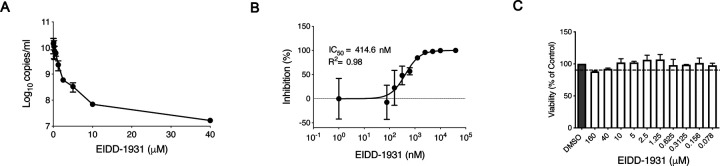
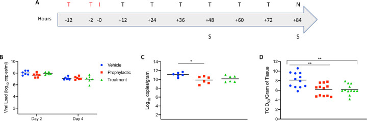
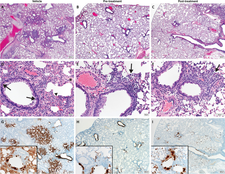
The COVID-19 pandemic progresses unabated in many regions of the world. An effective antiviral against SARS-CoV-2 that could be administered orally for use following high-risk exposure would be of substantial benefit in controlling the COVID-19 pandemic. Herein, we show that MK-4482, an orally administered nucleoside analog, inhibits SARS-CoV-2 replication in the Syrian hamster model. The inhibitory effect of MK-4482 on SARS-CoV-2 replication was observed in animals when the drug was administered either beginning 12 hours before or 12 hours following infection in a high-risk exposure model. These data support the potential utility of MK-4482 to control SARS-CoV-2 infection in humans following high-risk exposure as well as for treatment of COVID-19 patients.


Conflict of interest statement

Conflict of Interest Statement: The authors have declared that no conflict of interest exists.

Figures

Figure 1:
Figure 1:. EIDD-1931 inhibits SARS-CoV-2 replication in human lung epithelial Calu-3 cells.
Cells were pretreated for 1 hour with differing EIDD-1931 concentrations, followed by infection with SARS-CoV-2 at a MOI of 0.01 for 1 hour. After 1 hour, media was replaced, and cells were cultured in the presence of drug for 24 hours at 37°C in a 5% CO2 incubator. (A) Virus yield in the cell supernatant was measured by quantitative RT-PCR of clarified culture supernatant by using primer and probe sets to quantify total viral RNA (N gene; genomic and subgenomic RNA). (B) IC50 values were determined using results from the RT-PCR following log-based transformation of drug concentrations and normalization to percentage inhibition based on diluent alone controls by fitting to drug-dose response curves using Prism software. (C) Absence of toxicity (>90% viability; shown by dotted line) at highest EIDD-1931 concentration used for analysis of SARS-CoV-2 replication (40μM) was confirmed using CellTiter-Glo® 2.0 Assay (Promega, Corp., Madison, WI, USA) as per manufacturer’s protocol. For A to C, means are shown ± standard deviation.
Figure 2:
Figure 2:. Syrian hamster model - Study design, viral shedding, viral load, infectious titers and viral antigen.
(A) Study design. Hamsters were infected with SARS-CoV-2 by the intranasal route. MK-4482 was administered either pre-infection at 12 and 2 hours prior to infection, or post-infection with treatment started 12 hours post-infection. Treatment was then continued in both treatment groups every 12 hours for 3 consecutive days until end of the experiment. Animals were euthanized on day 4 and lungs were harvested for pathology and virology. ‘T’ denotes treatment (red: pre-infection and black: post-infection treatments); ‘I’ denotes infection; ‘S’ denotes swab samples and ‘N’ indicates necropsy. (B) Viral shedding. Oral swabs were collected on days 2 and 4 post-infection to measure viral shedding, determined by RT-PCR (N gene: genomic and subgenomic) (C) Viral load in lung tissue. Lung viral loads based on RT-PCR (N gene: genomic and subgenomic) were determined as a correlate for lower respiratory tract infection. (D) Infectious virus in lung tissue. Lung samples were homogenized and titered for infectious on Vero E6 cells. Infectious titers were determined as TCID50 equivalents using the Reed-Muench method. Two independent lung samples were measured from each animal. (B-D) Blue circle, vehicle control; red square, pre-infection treatment; green triangle, post-infection treatment. Summary of Results: (B) No statistical significance in virus shedding was found between either of the two MK-4482 treatment groups and vehicle controls. (C) There was a significant difference in lung viral loads between the pre-infection group compared to the vehicle control. Although the post-infection group trended towards lower levels, there was no significant difference between this group and vehicle control. (D) Infectious titers in the lungs were significantly different between both pre-infection and post-infection groups, compared to vehicle control group, but no significance was found between treatment groups from each other. For B to D geometric means are shown. ANOVA followed by Kruskal-Wallis analysis and a pairwise Wilcox test was used to analyze differences among groups. *p<0.05, **p<0.008
Figure 3:
Figure 3:. Pathological analysis of the lung tissue.
Hematoxylin and eosin (H&E) staining was used on lung sections to examine lung pathology post-inoculation. Immunohistochemistry (IHC) was used to detect viral antigen in the same lung sections. (A, D and G) untreated vehicle control, (B, E and H) pre-infection treatment with antiviral drug MK-4482 and (C, F and I) post-infection treatment with MK-4482. (A-F) H&E stain (G, H and I) IHC for SARS-CoV-2 nucleocapsid antibody. (A) Lung 20X: multifocal, moderate broncho-interstitial pneumonia. (B and C) Lung 20X: minimal peribronchial interstitial pneumonia. (D) Lung 200X epithelial cell necrosis (arrow), edema (asterisk), interstitial pneumonia (arrowhead). (E and F) peribronchial and interstitial infiltrates (arrow). (G) Lung 20X; insert 200X: numerous immunoreactive bronchiolar epithelial cells, type I and II pneumocytes and fewer macrophages. (H and I) Lung 20X; insert 200X: scattered to moderate numbers of immunoreactive bronchiolar epithelial cells, type I and II pneumocytes and macrophages.
Figure 4:
Figure 4:. Morphometric analysis of viral antigen and drug concentration in the lungs.
(A) A longitudinal cross section of the right lung was stained for viral antigen and scanned to measure the total amount of viral antigen present in the lung section. (B) EIDD-1931 concentrations in the lungs. (A and B) Blue circle, vehicle control; red square, pre-infection treatment; green triangle, post-infection treatment. Summary of results: (A) The area of lung staining positive for viral antigen showed a statistically significant difference between both of the MK-4482 treatment groups, compared to vehicle controls. No difference between individual treatment groups was present. For A and B, means are shown. ANOVA followed by Kruskal-Wallis analysis and a pairwise Wilcox test was used to analyze differences among groups. **p<0.008

References

    1. Lu R., et al. Genomic characterisation and epidemiology of 2019 novel coronavirus: implications for virus origins and receptor binding. Lancet 395, 565–574 (2020). - PMC - PubMed
    1. Chen N., et al. Epidemiological and clinical characteristics of 99 cases of 2019 novel coronavirus pneumonia in Wuhan, China: a descriptive study. Lancet 395, 507–513 (2020). - PMC - PubMed
    1. WHO. Coronavirus disease (COVID-2019) situation reports. Vol. 2020 (2020).
    1. Gibney E. Whose coronavirus strategy worked best? Scientists hunt most effective policies. Nature 581, 15–16 (2020). - PubMed
    1. Cowling B.J., et al. Impact assessment of non-pharmaceutical interventions against coronavirus disease 2019 and influenza in Hong Kong: an observational study. Lancet Public Health 5, e279–e288 (2020). - PMC - PubMed

Publication types