Review
. 2020 Nov 1;35(6):375-390.
doi: 10.1152/physiol.00008.2020.

The Pathophysiology of Rett Syndrome With a Focus on Breathing Dysfunctions

Affiliations
Review

The Pathophysiology of Rett Syndrome With a Focus on Breathing Dysfunctions

Jan-Marino Ramirez et al. Physiology (Bethesda). .

Abstract

Rett syndrome (RTT), an X-chromosome-linked neurological disorder, is characterized by serious pathophysiology, including breathing and feeding dysfunctions, and alteration of cardiorespiratory coupling, a consequence of multiple interrelated disturbances in the genetic and homeostatic regulation of central and peripheral neuronal networks, redox state, and control of inflammation. Characteristic breath-holds, obstructive sleep apnea, and aerophagia result in intermittent hypoxia, which, combined with mitochondrial dysfunction, causes oxidative stress-an important driver of the clinical presentation of RTT.

Keywords: autonomic dysregulation; breathing; dysphagia; oxidative stress.


Figures

FIGURE 1.
FIGURE 1.
The dynamic nature of RTT pathophysiology Schematic illustrating some of the hypothesized interactions that may underlie the RTT phenotype. RTT is associated with disturbances in a variety of CNS regions, resulting in a host of clinical phenotypes. These clinical presentations, in turn, have various pathophysiological consequences that themselves are dysregulated in RTT. This will further exaggerate or create additional symptoms. Important drivers of the RTT pathophysiology are mitochondrial and inflammatory dysfunctions. Targeting specific pathophysiological dysfunctions in RTT shows great therapeutic promise, yet this specific approach may not suffice to reestablish all the interacting dysregulations that are the hallmark of RTT.
FIGURE 2.
FIGURE 2.
Disruption of excitatory-inhibitory balance in Mecp2 KO (knockout) mouse model In normal, wild-type (WT) mice, the activity of excitatory and inhibitory neurons is delicately balanced, and the overall excitability of the entire network is tightly regulated. A: network diagrams illustrate the self-regulation of excitatory-inhibitory (E-I) balance in WT mice. Excitatory neurons (blue circles), inhibitory neurons (red circles), and their synapses (lines) are shown. Circle diameter indicates activity level, and line color indicates presynaptic identity. An example spiking trace from an excitatory neuron [in this case a L5 pyramidal neuron (35)] is shown. Normally, in WT animals, E-I is balanced. In cases in which a perturbation occurs, network activity levels may change, shown here by concomitant increase in E and I activity (middle). Homeostatic plasticity mechanisms work to return the global network state to a normal level of I-E activity and balance, although individual connections and cell activities may have changed. B: in Rett Syndrome, the balance is shifted toward hypoactivity and hypoconnectivity. This is shown as a reduction in excitatory activity (blue circle diameters) and number of synapses (lines). As in A above, an example trace of spiking activity from a L5 pyramidal neuron in a Mecp2 KO mouse is shown (35). The overall E-I balance is shifted to inhibition, and the network activity is decreased in the Mecp2 KO mouse; plasticity is impaired, and the network lacks the normal ability to achieve E-I balance or respond to perturbations normally. Potential therapeutic mechanisms are listed in the green shaded boxes. Administration of IGF-1 analogs (Trofenetide) and NMDA agonists (Ketamine) both not only restore EI balance in forebrain networks but additionally alleviate a number of clinical symptoms. An increase in excitatory postsynaptic current (EPSC) size under IGF-1 administration is one potential mechanism of E-I balance restoration, shown in the middle of the figure.
FIGURE 3.
FIGURE 3.
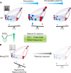
The dynamic interplay between breathholds and irregular breathing in RTT Hypothesized interactions are represented in this schematic. Episodic breathholds generated by disturbances in the Kolliker-Fuse (KF) regions of the pons lead to intermittent hypoxia, which may be a critical driver for irregular breathing. The effect of intermittent hypoxia is exaggerated by mitochondrial dysfunctions and reactive oxygen species (ROS) production. This disturbed REDOX regulation leads to carotid body hyperactivity, which, via an already disturbed nTS, will induce irregular breathing in the ventral respiratory group (medulla VRG). Right: each dot in the two graphs represents the irregularity score of an individual patient (RTT) or control subject. Note that not every patient with RTT shows irregular breathing (pink shaded box), and many RTT patients have irregularity scores similar to those of controls (green shaded boxes). Normal breathing is maintained by homeostatic mechanisms. Graphs and traces modified from Refs. , , with permission from Pediatric Pulmonology and Pediatric Research, respectively.
FIGURE 4.
FIGURE 4.
Tracings and anatomic locations depicting normal and discoordinated swallow-breathing behavior A: muscle electromyograms (EMG) traces and pressure measurements depict normal sequential swallow pattern in the healthy adult rat with the colors coordinated to different muscles throughout the aerodigestive tract. B: anatomic depiction of the aerodigestive tract involved in both breathing and swallow. C: swallow-breathing coordination in a healthy human with spirometry airflow trace in pink (downward deflection indicating inspiration, and upward indicating expiration) with zero airflow, along with surface EMG submental complex activation (orange), indicating swallow. The leading complex (mylohyoid and geniohyoid in rat) or submental complex in the human indicates the oral phase (orange) and correlates to the anatomic structure listed in orange (B). The thyroarytenoid and thyropharyngeus in blue (A) measures the pharyngeal phase of swallow listed in blue (B). The esophageal pressure, red (A) depicts the esophageal phase and is the last in the swallow sequence. Activation of the diaphragm (in pink) during swallow occurs during leading complex activation. D: bolus travel in normal swallow (red arrow) bolus travels from pharynx to esophagus. E: spirometry (pink) and respiratory inductive plethysmography bands (purple) around the chest and abdomen show swallow-breathing coordination in a patient with Rett syndrome. Normal eupnea occurs until the spoon comes in contact with the patient; lack of airflow and no movement in the chest or abdomen represent the apneic period; and swallow occurrence at the end is depicted by the flat line at the end of the feeding session, followed by normal eupnea. Dysphagic swallow, penetration (green arrow) of the bolus passes to the larynx but does not go below the vocal folds, whereas in more severe aspiration (blue) the bolus passes through the vocal folds and into the trachea leading to the lungs. D and E are modified from Ref. , with permission from Developmental Medicine and Child Physiology.

References

    1. Abdala AP, Bissonnette JM, Newman-Tancredi A. Pinpointing brainstem mechanisms responsible for autonomic dysfunction in Rett syndrome: therapeutic perspectives for 5-HT1A agonists. Front Physiol 5: 205, 2014. doi:10.3389/fphys.2014.00205. - DOI - PMC - PubMed
    1. Abdala AP, Dutschmann M, Bissonnette JM, Paton JF. Correction of respiratory disorders in a mouse model of Rett syndrome. Proc Natl Acad Sci USA 107: 18208–18213, 2010. doi:10.1073/pnas.1012104107. - DOI - PMC - PubMed
    1. Abdala AP, Toward MA, Dutschmann M, Bissonnette JM, Paton JF. Deficiency of GABAergic synaptic inhibition in the Kölliker-Fuse area underlies respiratory dysrhythmia in a mouse model of Rett syndrome. J Physiol 594: 223–237, 2016. doi:10.1113/JP270966. - DOI - PMC - PubMed
    1. Abraham SS, Taragin B, Djukic A. Co-occurrence of dystonic and dyskinetic tongue movements with oral apraxia in post-regression dysphagia in classical Rett syndrome years of life 1 through 5. Dysphagia 30: 128–138, 2015. doi:10.1007/s00455-014-9587-9. - DOI - PubMed
    1. Amaddeo A, De Sanctis L, Arroyo JO, Khirani S, Bahi-Buisson N, Fauroux B. Polysomnographic findings in Rett syndrome. Eur J Paediatr Neurol 23: 214–221, 2019. doi:10.1016/j.ejpn.2018.09.003. - DOI - PubMed

Publication types