

Point-of-care Lung Ultrasound Is Useful to Evaluate Emergency Department Patients for COVID-19
- PMID: 33052813
- PMCID: PMC7673866
- DOI: 10.5811/westjem.2020.8.49205
Point-of-care Lung Ultrasound Is Useful to Evaluate Emergency Department Patients for COVID-19
Abstract
Introduction: Coronavirus disease 2019 (COVID-19) can be a life-threatening lung disease or a trivial upper respiratory infection depending on whether the alveoli are involved. Emergency department (ED) evaluation of symptomatic patients with normal vital signs is frequently limited to chest auscultation and oro-nasopharyngeal swabs. We tested the null hypothesis that patients being screened for COVID-19 in the ED with normal vital signs and without hypoxia would have a point-of-care lung ultrasound (LUS) consistent with COVID-19 less than 2% of the time.
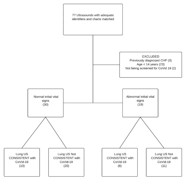
Methods: We performed a retrospective, structured, blinded ultrasound review and chart review in patients 14 years or older with symptoms prompting ED evaluation for COVID-19. We excluded those with known congestive heart failure or other chronic lung conditions likely to cause excessive B-lines on LUS. We used a two-sided exact hypothesis test for binomial random variables. We measured LUS diagnostic performance using computed tomography as the gold standard.
Results: We reviewed 77 charts; 49 met inclusion criteria. Vital signs were normal in 30/49 patients; 10 (33%) of these patients had LUS consistent with viral pneumonitis. We rejected the null hypothesis (p-value <0.001). The treating physicians' interpretations of their own point-of-care LUS had a sensitivity of 100% (95% confidence interval (CI), 74%, 100%), specificity 88% (95% CI, 47%, 100%), likelihood ratio (LR) positive of 5.8 (95% CI, 1.3, 25), and LR negative of 0.05 (95% CI, 0.03, 0.71) when compared to CT findings.
Conclusion: LUS had a meaningful detection rate for pneumonitis in symptomatic ED patients with normal vital signs who were being evaluated for COVID-19. We recommend at least LUS be used in addition to polymerase chain reaction testing when evaluating symptomatic ED patients for COVID-19.
Conflict of interest statement
Figures
Update of
-
Point of care lung ultrasound is useful when screening for CoVid-19 in Emergency Department patients.medRxiv [Preprint]. 2020 Jun 12:2020.06.09.20123836. doi: 10.1101/2020.06.09.20123836. medRxiv. 2020. Update in: West J Emerg Med. 2020 Sep 28;21(6):24-31. doi: 10.5811/westjem.2020.8.49205. PMID: 32587987 Free PMC article. Updated. Preprint.
Similar articles
-
Point of care lung ultrasound is useful when screening for CoVid-19 in Emergency Department patients.medRxiv [Preprint]. 2020 Jun 12:2020.06.09.20123836. doi: 10.1101/2020.06.09.20123836. medRxiv. 2020. Update in: West J Emerg Med. 2020 Sep 28;21(6):24-31. doi: 10.5811/westjem.2020.8.49205. PMID: 32587987 Free PMC article. Updated. Preprint.
-
Lung ultrasound in the emergency department - a valuable tool in the management of patients presenting with respiratory symptoms during the SARS-CoV-2 pandemic.BMC Emerg Med. 2020 Dec 7;20(1):96. doi: 10.1186/s12873-020-00389-w. BMC Emerg Med. 2020. PMID: 33287732 Free PMC article.
-
Point-of-care ultrasound (PoCUS) in the early diagnosis of novel coronavirus 2019 disease (COVID-19) in a first-level emergency department during a SARS-CoV-2 outbreak in Italy: a real-life analysis.Intern Emerg Med. 2022 Jan;17(1):193-204. doi: 10.1007/s11739-021-02643-w. Epub 2021 Apr 21. Intern Emerg Med. 2022. PMID: 33881727 Free PMC article.
-
Point-of-care lung ultrasound for the assessment of pneumonia: a narrative review in the COVID-19 era.J Med Ultrason (2001). 2021 Jan;48(1):31-43. doi: 10.1007/s10396-020-01074-y. Epub 2021 Jan 13. J Med Ultrason (2001). 2021. PMID: 33438132 Free PMC article. Review.
-
Thoracic imaging tests for the diagnosis of COVID-19.Cochrane Database Syst Rev. 2020 Sep 30;9:CD013639. doi: 10.1002/14651858.CD013639.pub2. Cochrane Database Syst Rev. 2020. Update in: Cochrane Database Syst Rev. 2020 Nov 26;11:CD013639. doi: 10.1002/14651858.CD013639.pub3. PMID: 32997361 Updated.
Cited by
-
M-BLUE protocol for coronavirus disease-19 (COVID-19) patients: interobserver variability and correlation with disease severity.Clin Radiol. 2021 May;76(5):379-383. doi: 10.1016/j.crad.2021.02.003. Epub 2021 Feb 17. Clin Radiol. 2021. PMID: 33663912 Free PMC article.
-
The utility of lung ultrasound in COVID-19: A systematic scoping review.Ultrasound. 2020 Nov;28(4):208-222. doi: 10.1177/1742271X20950779. Epub 2020 Aug 17. Ultrasound. 2020. PMID: 36959895 Free PMC article.
-
A Comparison of Lung Ultrasound and Computed Tomography in the Diagnosis of Patients with COVID-19: A Systematic Review and Meta-Analysis.Diagnostics (Basel). 2021 Jul 27;11(8):1351. doi: 10.3390/diagnostics11081351. Diagnostics (Basel). 2021. PMID: 34441286 Free PMC article. Review.
-
Point-of-care lung ultrasound predicts hyperferritinemia and hospitalization, but not elevated troponin in SARS-CoV-2 viral pneumonitis in children.Sci Rep. 2024 Mar 11;14(1):5899. doi: 10.1038/s41598-024-55590-9. Sci Rep. 2024. PMID: 38467670 Free PMC article.
-
Review of COVID-19 testing and diagnostic methods.Talanta. 2022 Jul 1;244:123409. doi: 10.1016/j.talanta.2022.123409. Epub 2022 Mar 31. Talanta. 2022. PMID: 35390680 Free PMC article. Review.
References
-
-
Ref 3 the journal abbreviation is Wang Z, Yang B, Li Q, Wen L, Zhang R. Clinical features of 69 cases with coronavirus disease 2019 in Wuhan, China. Clin Infect Dis. 2020;71(15):769–77.
-
MeSH terms
Grants and funding
LinkOut - more resources
Full Text Sources
Medical
Miscellaneous