Phase-Aberration Correction for HIFU Therapy Using a Multielement Array and Backscattering of Nonlinear Pulses
- PMID: 33052845
- PMCID: PMC8476183
- DOI: 10.1109/TUFFC.2020.3030890
Phase-Aberration Correction for HIFU Therapy Using a Multielement Array and Backscattering of Nonlinear Pulses
Abstract
Phase aberrations induced by heterogeneities in body wall tissues introduce a shift and broadening of the high-intensity focused ultrasound (HIFU) focus, associated with decreased focal intensity. This effect is particularly detrimental for HIFU therapies that rely on shock front formation at the focus, such as boiling histotripsy (BH). In this article, an aberration correction method based on the backscattering of nonlinear ultrasound pulses from the focus is proposed and evaluated in tissue-mimicking phantoms. A custom BH system comprising a 1.5-MHz 256-element array connected to a Verasonics V1 engine was used as a pulse/echo probe. Pulse inversion imaging was implemented to visualize the second harmonic of the backscattered signal from the focus inside a phantom when propagating through an aberrating layer. Phase correction for each array element was derived from an aberration-correction method for ultrasound imaging that combines both the beamsum and the nearest neighbor correlation method and adapted it to the unique configuration of the array. The results were confirmed by replacing the target tissue with a fiber-optic hydrophone. Comparing the shock amplitude before and after phase-aberration correction showed that the majority of losses due to tissue heterogeneity were compensated, enabling fully developed shocks to be generated while focusing through aberrating layers. The feasibility of using a HIFU phased-array transducer as a pulse-echo probe in harmonic imaging mode to correct for phase aberrations was demonstrated.
Figures


 , second harmonic
  and third harmonic
  at a system drive voltage of 8V (68 W acoustic power) [46].


 represents the length of the focal lobe of the second harmonic. Bottom: beamsum around the focus.
 . The area between the two vertical dashed lines illustrates a cluster of HIFU array elements with low correlation coefficients to the beamsum signal, and a corresponding high variance in phase error estimates.
 , plotted together with the output of Step 2 (beamsum method)
  . High phase error variance from the beamsum method was successfully removed by the “anchored” nearest neighbor method.
 .
.
 .
.
 , with the aberration-inducing phantom and correction:
  , in water:
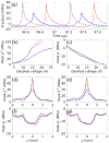
  . (a) Waveforms at the focus for the same derated voltage of 15V in water. (b) Peak positive pressure and (c) peak negative pressure at the focus as functions of the input voltage (derated if the phantom is present). (d)-(g) Transverse beam profiles in the focal plane for the same derated voltage as 15V in water.
References
-
- ter Haar G and Coussios C, “High intensity focused ultrasound: physical principles and devices,” International Journal of Hyperthermia. vol. 23, no. 2, pp. 89–104, 2007. - PubMed
Publication types
MeSH terms
Grants and funding
LinkOut - more resources
Full Text Sources
Other Literature Sources

