Review
. 2020 Oct 1;59(Suppl4):iv6-iv17.
doi: 10.1093/rheumatology/keaa250.

Classification vs diagnostic criteria: the challenge of diagnosing axial spondyloarthritis

Affiliations
Review

Classification vs diagnostic criteria: the challenge of diagnosing axial spondyloarthritis

Denis Poddubnyy. Rheumatology (Oxford). .

Abstract

In recent years, significant progress has been made in improving the early diagnosis of spondyloarthritides (SpA), including axial SpA. Nonetheless, there are still issues related to the application of classification criteria for making the primary diagnosis of SpA in the daily practice. There are substantial conceptional and operational differences between the diagnostic vs classification approach. Although it is not possible to develop true diagnostic criteria for natural reasons as discussed in this review, the main principles of the diagnostic approach can be clearly defined: consider the pre-test probability of the disease, evaluate positive and negative results of the diagnostic test, exclude other entities, and estimate the probability of the disease at the end. Classification criteria should only be applied to patients with an established diagnosis and aimed at the identification of a rather homogeneous group of patients for the conduction of clinical research.

Keywords: ankylosing spondylitis; axial spondyloarthritis; classification; diagnosis; magnetic resonance imaging.


Figures

<sc>Fig</sc>. 1
Fig. 1
ASAS classification criteria for axial and peripheral spondyloarthritis (A) ASAS classification criteria for axial spondyloarthritis; (B) ASAS classification criteria for peripheral spondyloarthritis. *Sacroiliitis on imaging refers to definite radiographic sacroiliitis according to the modified New York criteria or active sacroiliitis on MRI according to the ASAS definition. **Peripheral arthritis: usually predominantly lower limbs and/or asymmetric arthritis; enthesitis: clinically assessed; dactylitis: clinically assessed. SpA: spondyloarthritis.
<sc>Fig</sc>. 2
Fig. 2
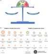
The diagnostic scale and weights of diagnostic test results for axial spondyloarthritis The balanced scale (A) indicates an undetermined diagnostic situation, in which other tests are normally needed. Positive and negative test results depending on their weights would change the balance towards more or less likely diagnosis. The weights of the positive and negative diagnostic tests (B) reflected by the size of the balls represent examples based on approximation of the literature data [8]. The real diagnostic weight might be different depending on the region, background population, and referral structures – see article text for further details. An alternative, more likely explanation of symptoms (differential diagnosis), is attributed to the weights of negative test results. SpA: spondyloarthritis.
<sc>Fig</sc>. 3
Fig. 3
Conventional radiography, MRI-STIR, MRI-T1 of sacroiliac joints, and a diagnostic scale for patient A A 55-year-old male patient referred by an ophthalmologist because of an episode of acute anterior uveitis and inflammatory back pain of intermittent intensity for about 15 to 20 years. HLA-B27 is positive, CRP is normal. In addition to some suspicious radiographic changes, MRI of sacroiliac joints showed definite active inflammatory and structural changes compatible with SpA. A diagnosis of axial SpA was made. See the article text for further details. SpA: spondyloarthritis; STIR: short tau inversion recovery.
<sc>Fig</sc>. 4
Fig. 4
Conventional radiography, MRI-STIR, MRI-T1 of sacroiliac joints, and a diagnostic scale for patient B A 53-year-old male patient referred by an ophthalmologist because of two recent episodes of acute anterior uveitis and inflammatory back pain of intermittent intensity for about 20 years. HLA-B27 is positive, CRP is normal. Conventional radiography of sacroiliac joints showed suspicious changes, but no SpA-compatible changes could be found on MRI. At the same time, degenerative changes of the intervertebral disc represent the most likely explanation of back pain in this case. See the article text for further details. SpA: spondyloarthritis; STIR: short tau inversion recovery.

Similar articles

Cited by

References

    1. Sieper J, Poddubnyy D. Axial spondyloarthritis. Lancet 2017;390:73–84. - PubMed
    1. Proft F, Poddubnyy D. Ankylosing spondylitis and axial spondyloarthritis: recent insights and impact of new classification criteria. The Adv Musculoskelet Dis 2018;10:129–39. - PMC - PubMed
    1. van der Linden S, Valkenburg HA, Cats A. Evaluation of diagnostic criteria for ankylosing spondylitis. A proposal for modification of the New York criteria. Arthritis Rheum 1984;27:361–8. - PubMed
    1. Boel A, Molto A, van der Heijde D et al. Do patients with axial spondyloarthritis with radiographic sacroiliitis fulfil both the modified New York criteria and the ASAS axial spondyloarthritis criteria? Results from eight cohorts. Ann Rheum Dis 2019;78:1545–9. - PubMed
    1. Redeker I, Callhoff J, Hoffmann F et al. Determinants of diagnostic delay in axial spondyloarthritis: an analysis based on linked claims and patient-reported survey data. Rheumatology 2019;58:1634–8. - PubMed

Publication types

MeSH terms