. 2020 Dec:146:105131.
doi: 10.1016/j.nbd.2020.105131. Epub 2020 Oct 11.

The SARS-CoV-2 spike protein alters barrier function in 2D static and 3D microfluidic in-vitro models of the human blood-brain barrier

Affiliations

The SARS-CoV-2 spike protein alters barrier function in 2D static and 3D microfluidic in-vitro models of the human blood-brain barrier

Tetyana P Buzhdygan et al. Neurobiol Dis. 2020 Dec.

Abstract

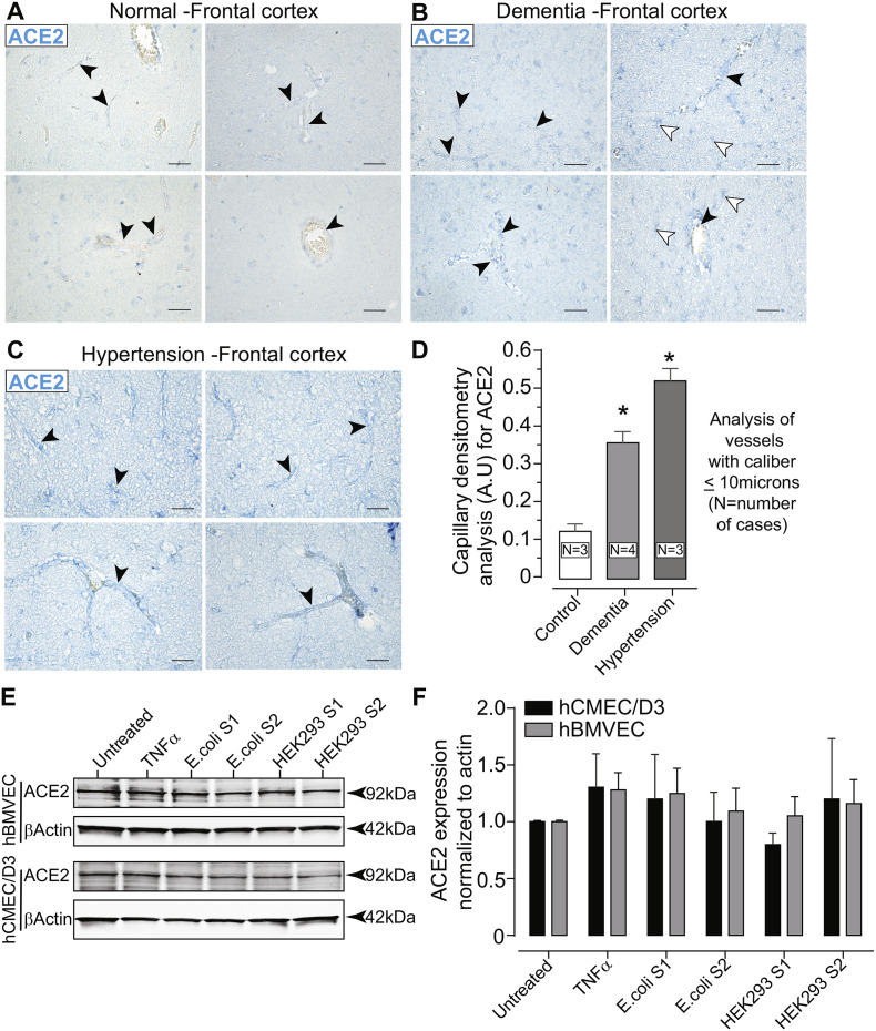
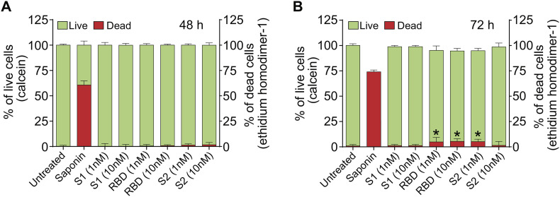
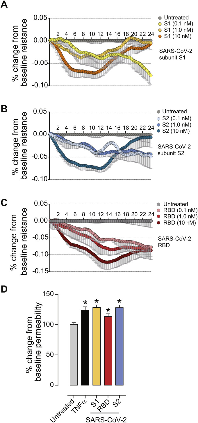
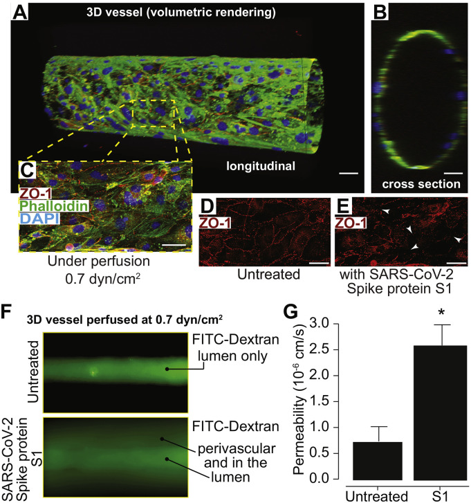
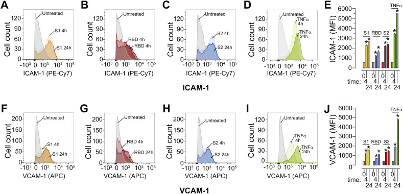
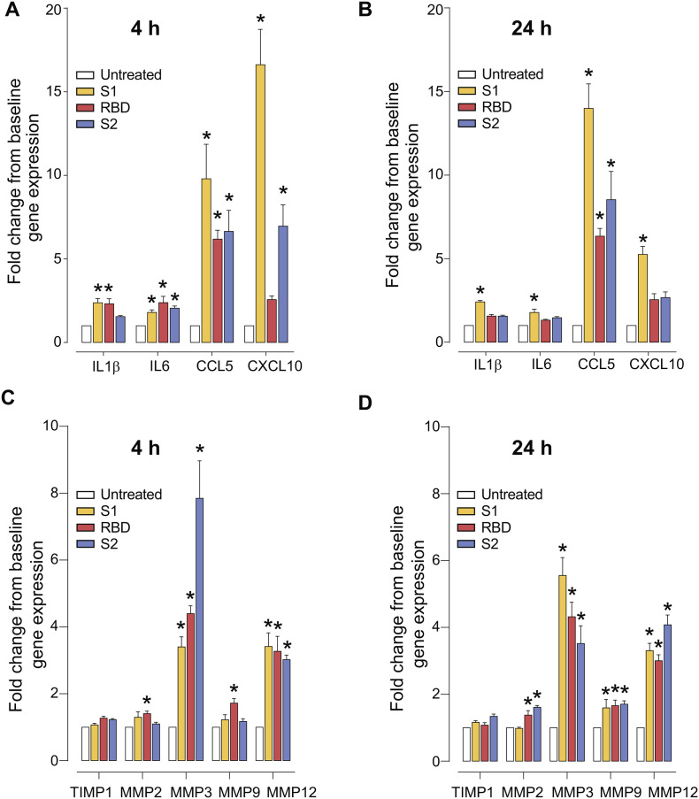
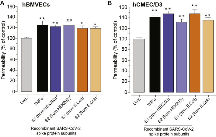
As researchers across the globe have focused their attention on understanding SARS-CoV-2, the picture that is emerging is that of a virus that has serious effects on the vasculature in multiple organ systems including the cerebral vasculature. Observed effects on the central nervous system include neurological symptoms (headache, nausea, dizziness), fatal microclot formation and in rare cases encephalitis. However, our understanding of how the virus causes these mild to severe neurological symptoms and how the cerebral vasculature is impacted remains unclear. Thus, the results presented in this report explored whether deleterious outcomes from the SARS-CoV-2 viral spike protein on primary human brain microvascular endothelial cells (hBMVECs) could be observed. The spike protein, which plays a key role in receptor recognition, is formed by the S1 subunit containing a receptor binding domain (RBD) and the S2 subunit. First, using postmortem brain tissue, we show that the angiotensin converting enzyme 2 or ACE2 (a known binding target for the SARS-CoV-2 spike protein), is ubiquitously expressed throughout various vessel calibers in the frontal cortex. Moreover, ACE2 expression was upregulated in cases of hypertension and dementia. ACE2 was also detectable in primary hBMVECs maintained under cell culture conditions. Analysis of cell viability revealed that neither the S1, S2 or a truncated form of the S1 containing only the RBD had minimal effects on hBMVEC viability within a 48 h exposure window. Introduction of spike proteins to invitro models of the blood-brain barrier (BBB) showed significant changes to barrier properties. Key to our findings is the demonstration that S1 promotes loss of barrier integrity in an advanced 3D microfluidic model of the human BBB, a platform that more closely resembles the physiological conditions at this CNS interface. Evidence provided suggests that the SARS-CoV-2 spike proteins trigger a pro-inflammatory response on brain endothelial cells that may contribute to an altered state of BBB function. Together, these results are the first to show the direct impact that the SARS-CoV-2 spike protein could have on brain endothelial cells; thereby offering a plausible explanation for the neurological consequences seen in COVID-19 patients.

Keywords: Blood-brain barrier; COVID-19; Cerebral vascular biology; Microfluidic chip; Neuroinflammation; SARS-CoV-2; Tissue engineering.


Conflict of interest statement

The authors declare no competing interests.

Figures

Fig. 1
Fig. 1
ACE2 is expressed on the cerebral vasculature and in primary human brain microvascular endothelial cells (hBMVECs). Paraffin-embedded brain tissue was sectioned at 5 μm and immunostained for ACE2. Representative images of the frontal cortex from cases with no abnormal neuropathology (control) (A), dementia (B) and hypertension (C). The images were scanned at 40× objective magnification with ACE2 expression shown in blue (Vector Blue). Black arrow heads indicate the vascular presentation of ACE2 expression in various caliber vessels. White arrow heads point to ACE2 expression in parenchymal cells. Scalebars = 25 μm. (D) Bar graph of the quantification for ACE2 expression in capillary sized vessels (under 10 μm in diameter/caliber). (E) Western blots of hBMVECs and hCMEC/D3 cell lysates probed with ACE2 antibodies after cells were exposed to 10 nM of the SARS-CoV-2 subunit S1 or subunit S2 expressed either in E.coli or in HEK293 cells. (F) Bar graph of densitometry of the ACE2 immunoblot normalized to β-Actin. The experiment using hBMVECs was performed with three different donors and repeated three times. No significant differences between the groups were observed. (For interpretation of the references to colour in this figure legend, the reader is referred to the web version of this article.)
Fig. 2
Fig. 2
SARS-CoV-2 spike protein does not affect brain endothelial cell viability. hBMVECs were treated with 1 nM and 10 nM of the SARS-CoV-2 subunit S1, SARS-CoV-2 RBD, and SARS-CoV-2 subunit S2 for 48 h (A) and 72 h (B). Cell viability was determined using the Live/Dead Cytotoxicity assay. Calcein positive (green) indicates live cells while ethidium homodimer-1 (EthD-1, red) indicates dead cells. Saponin was used a positive control. Data represents the ratio of live or dead cells in the total cell number and was obtained from two different donors, each performed in 6 replicates. Results are presented as mean ± SEM, n = 12, *p < 0.05. p-values were computed using one-way ANOVA and Tukey post-hoc test. (For interpretation of the references to colour in this figure legend, the reader is referred to the web version of this article.)
Fig. 3
Fig. 3
SARS-CoV-2 spike protein compromises endothelial barrier properties. (A-C) Barrier electrical resistance was modelled based on continuous cell-substrate impedance readings recorded at 6 frequencies (400 Hz – 48 kHz) every 6 min for the duration of the time shown. Endothelial monolayers were treated with 0.1 nM, 1 nM or 10 nM of SARS-CoV-2 subunit S1, SARS-CoV-2 RBD, SARS-CoV-2 subunit S2 or left untreated to serve as a baseline. Treatments were initiated at 0 timepoint. The experiment was performed in quadruplicates and repeated three times using primary cells obtained from three different donors. Each data point is represented as the percentage of change from the baseline (mean ± SEM), n = 12. D. Barrier permeability to a small molecular tracer was determined 1 h after treatment using the 3 kDa FITC-conjugated DEAE-dextran. Endothelial monolayers were treated with 100 ng/mL TNF-α, 10 nM of SARS-CoV-2 subunit S1, SARS-CoV-2 RBD, SARS-CoV-2 subunit S2 or left untreated to serve as a baseline. The experiment was performed in quadruplicates and repeated three times using primary cells obtained from three different donors. Each data point is represented as mean ± SEM, n = 12, p-values were computed using one-way ANOVA and Tukey post-hoc test.
Fig. 4
Fig. 4
SARS-CoV-2 subunit S1 alters barrier status in a 3D tissue engineered microfluidic model of the human BBB. Confocal microscopy and volumetric rendering were used to visualize the tissue engineered vessel. (A) Shows a longitudinal view of an endothelialized void after perfusion that formed a predictive vessel geometry analogous to those found within the brain. (B) provides a cross sectional perspective indicating a single layer of endothelial cells. In (C) a representative merged image of the engineered vessel constructs fixed and immunestained for the tight junction protein, ZO-1, along with phalloidin to label actin and the nuclear stain, DAPI. (D) shows the typical ZO-1 membranous pattern expected in mature barrier forming brain endothelial cells. (E) after perfusion for 2 h of SARS-CoV-2 subunit S1 (10 nM), constructs were also fixed and immunolabeled for ZO-1. The arrows point to areas in which the ZO-1 cellular pattern is discontinuous, punctate or absent signifying areas of barrier breach. Scalebar = 20 μm. (F) Fluorescence intensity after ten minutes of perfusion with 4 kDa FITC-dextran, indicating the impaired barrier function in vessels perfused after 2 h of the S1 spike protein versus untreated controls. G) Quantitative measurements for permeability coefficients of vessels exposed to the SARS-CoV-2 subunit S1 compared to untreated controls. Data was analyzed using Kruskal-Willis test, n = 3, *p < 0.05.
Fig. 5
Fig. 5
SARS-CoV-2 spike protein triggers enhanced surface expression of adhesion molecules. Human brain microvascular endothelial cells (hBMVECs) were treated with 10 nM of SARS-CoV-2 subunit S1, SARS-CoV-2 RBD, SARS-CoV-2 subunit S2, or 100 ng/mL of TNF-α for 4 h or 24 h. Cells were stained for ICAM-1 and VCAM-1 expression and analyzed using a FACS Canto II flow cytometer. Shown are representative histograms for ICAM-1 expression in response to SARS-CoV-2 subunit S1 (A), SARS-CoV-2 RBD (B), SARS-CoV-2 subunit S2 (C), TNF-α (D) and the bar graph quantification of the Mean Fluorescent Intensity (MFI) (E). Representative histogram for VCAM-1 expression in response to SARS-CoV-2 subunit S1 (F), SARS-CoV-2 RBD (G), SARS-CoV-2 subunit S2 (H), TNF-α (I) and the bar graph quantification of MFI (J), n = 3, *p < 0.05.
Fig. 6
Fig. 6
SARS-CoV-2 spike protein triggers pro-inflammatory responses and upregulation of MMPs in hBMVECs. Confluent hBMVEC monolayers were incubated with 10 nM of SARS-CoV-2 subunit S1, SARS-CoV-2 RBD, SARS-CoV-2 subunit S2, for time indicated or left untreated to serve as a baseline control. Target cytokine genes analyzed included: IL1β, IL6, CCL5, CXCL10 at 4 h (A) and 24 h (B). Gene expression analysis for MMP2, MMP3, MMP9, MMP12 and the MMP inhibitor TIMP1 are shown for 4 h (C) and 24 h (D) respectively. Experiments were performed in quadruplicates and repeated three times using primary cells obtained from three different donors. Each bar represents a fold-change mean ± SEM, n = 12. Data sets were analyzed using one-way ANOVA and post-hoc comparison to the untreated condition were computed using Tukey post-hoc test with *p < 0.05.
Supplementary Fig. S1
The following are the supplementary data related to this article.Supplementary Fig. S1
SARS-CoV-2 spike protein triggers an increase in paracellular permeability in hCMEC/D3 monolayers.

Update of

References

    1. Ackermann M., et al. Pulmonary vascular Endothelialitis, thrombosis, and angiogenesis in Covid-19. N. Engl. J. Med. 2020 doi: 10.1056/NEJMoa2015432. - DOI - PMC - PubMed
    1. Adamson R.H., Lenz J.F., Curry F.E. Quantitative laser scanning confocal microscopy on single capillaries: permeability measurement. Microcirculation. 1994;1:251–265. doi: 10.3109/10739689409146752. - DOI - PubMed
    1. Al-Obaidi M.M.J., et al. Disruption of the blood brain barrier is vital property of neurotropic viral infection of the central nervous system. Acta Virol. 2018;62:16–27. doi: 10.4149/av_2018_102. - DOI - PubMed
    1. Andrews A.M., et al. Characterization of human fetal brain endothelial cells reveals barrier properties suitable for in vitro modeling of the BBB with syngenic co-cultures. J. Cereb. Blood Flow Metab. 2018;38:888–903. doi: 10.1177/0271678X17708690. - DOI - PMC - PubMed
    1. Berger J.R., Avison M. The blood brain barrier in HIV infection. Front. Biosci. 2004;9:2680–2685. doi: 10.2741/1427. - DOI - PubMed

Publication types

MeSH terms