Case Reports
. 2020 Oct 12;8(4):117.
doi: 10.3390/dj8040117.

Superficial Temporal Artery Perforator Flap: Indications, Surgical Outcomes, and Donor Site Morbidity

Affiliations
Case Reports

Superficial Temporal Artery Perforator Flap: Indications, Surgical Outcomes, and Donor Site Morbidity

Raffaele Rauso et al. Dent J (Basel). .

Abstract

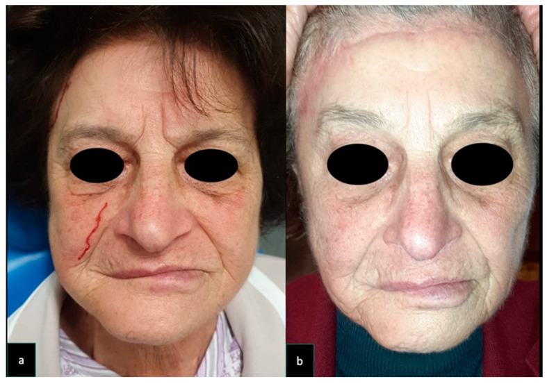
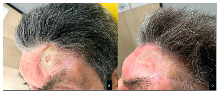
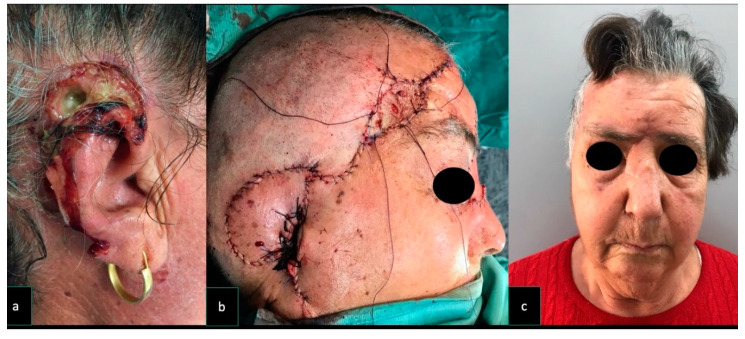
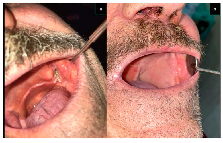
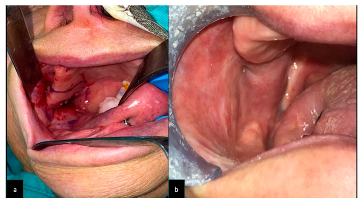
The aim of this retrospective case series was to discuss indications, surgical outcomes, and donor site morbidity in the use of superficial temporal artery perforator (STAP) flaps in intra-oral or extra-oral facial reconstruction. This study involved 9 patients treated with a STAP flap at the Maxillo-Facial Surgery Unit of the University of Campania "Luigi Vanvitelli", Naples. A STAP flap was used alone or in combination with other local flaps, for the coverage of facial soft tissue defects, after the resection of craniofacial malignant tumors (n = 7) or as a salvage flap, in partial or total microvascular flap loss (n = 2). The STAP flap was proven to be a valuable surgical option despite it not being frequently used in facial soft tissue reconstruction nor was it chosen as the first surgical option in patients under 70 year's old. Donor site morbidity is one of the major reasons why this flap is uncommon. Appropriate patient selection, surgical plan, and post-surgical touch-ups should be performed in order to reduce donor site scar morbidity.

Keywords: donor site morbidity; free flap combination; intraoral defect; reconstructive surgical procedures; superficial temporal artery island flap.


Conflict of interest statement

The authors declare no conflict of interest.

Figures

Figure 1
Figure 1
Case 6. A 79-year old woman underwent surgical excision for a recurrent left tuber maxillae squamous cell carcinoma. The patient had previously undergone a homolateral neck dissection and radiation therapy resulting in a depleted vessel neck. For this reason, a pedicle superficial temporal artery flap (9 × 9 cm) was raised and used to reconstruct the oral defect. The flap donor site was repaired with a skin graft and to reduce donor site scar morbidity, a forehead lift was implemented. (a) Pre-operative facial appearance of the patient. (b) Post-operative facial appearance of the patient. In order to reduce donor site morbidity and to achieve a symmetrical post-operative appearance of the forehead, a forehead lifting was performed.
Figure 2
Figure 2
Case 3. A 74 -year old man underwent surgical excision for a recurrent left tuber maxillae squamous cell carcinoma. The patient previously underwent a homolateral neck dissection and radiation therapy, resulting in a depleted vessel neck. For these reasons, a pedicle superficial temporal artery flap (7 × 7 cm) was raised and used to reconstruct the oral defect. The flap donor site was repaired with a skin graft. Five sessions of a 20 mg/mL cross-linked hyaluronic acid (HA) filler (Hyamira Basic, Nyuma pharma®, Arona, Italy) was injected into the grafted area. About 0.2 mL of HA per session was injected over a total of 5 sessions, spaced 30 days apart. After the HA was injected into the graft site, a better skin quality with a more elastic tissue, and a less depressed appearance of the area were noticed. (a) The concave appearance of the grafted site was improved, following several sessions of cross-linked HA injections (b).
Figure 3
Figure 3
Case 2. A 76-year old woman underwent excisions for a squamous cell carcinoma involving the external ear and mastoid area A pedicle superficial temporal artery flap (9 × 7 cm) was raised and used to reconstruct the ear defect. The flap donor site was repaired with a skin graft and a local advancement flap was harvested to reduce the skin graft size. At 6-months follow-up, the patient was pleased with the aesthetic result and was disease free. (a) Pre-operative appearance of a cancer involving external ear and mastoid area. (b) The reconstruction performed with STAP flap; the donor site was skin grafted. (c) Post-operative symmetric appearance of the brows at rest.
Figure 4
Figure 4
Same patient as Figure 2 (Case 3) (a) Intra-oral aspect of the cancer involving left tuber maxillae and cheek. The reconstruction performed with STAP flap (b).
Figure 5
Figure 5
Same patient as Figure 1 (Case 6) (a) Intra-oral aspect of the cancer involving right tuber maxillae and cheek. (b) The reconstruction performed with STAP flap.

References

    1. Mustarde J.C. Cheek rotation skin flap to the lower eyelid. In: Strauch B., editor. Grabb’s Encyclopedia of Flaps. Little Brown; Boston, MA, USA: 1990. p. 51.
    1. Dolen U.C., Baltu Y., Aydin O. Subunit Reconstruction of Mid-Facial Defects With Free Style Facial Perforator Flaps. J. Craniofac. Surg. 2018;29:1574–1577. doi: 10.1097/SCS.0000000000004535. - DOI - PubMed
    1. Wong C.H., Wei F.C. Microsurgical free flap in head and neck reconstruction. Head Neck. 2010;32:1236–1245. doi: 10.1002/hed.21284. - DOI - PubMed
    1. Esposito L., Razzano S., Lo Faro C., Dell’Aversana Orabona G., Schonauer F. Pinna fillet flap after advanced external ear tumor resection. JPRAS Open. 2016;8:9–13. doi: 10.1016/j.jpra.2016.02.002. - DOI
    1. Massarelli O., Vaira L.A., Gobbi R., Dell’aversana Orabona G., De Riu G. Reconstruction of full-thickness cheek defect with chimeric facial artery free flap: A case report. Microsurgery. 2018;38:427–431. doi: 10.1002/micr.30282. - DOI - PubMed

Publication types

LinkOut - more resources