Review
. 2020 Oct 12;21(20):7513.
doi: 10.3390/ijms21207513.

Functions of Osteocalcin in Bone, Pancreas, Testis, and Muscle

Affiliations
Review

Functions of Osteocalcin in Bone, Pancreas, Testis, and Muscle

Toshihisa Komori. Int J Mol Sci. .

Abstract

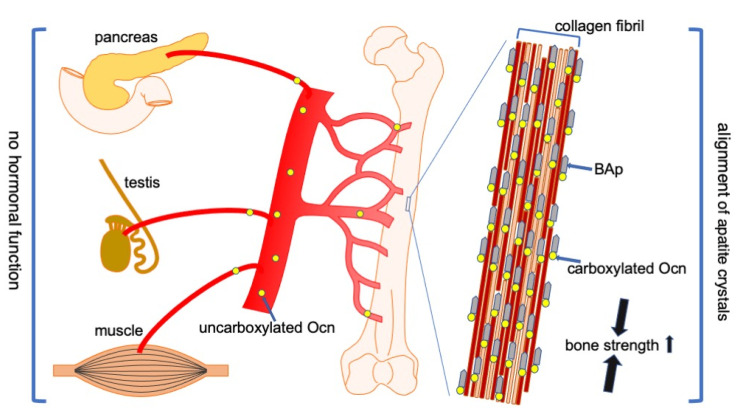
Osteocalcin (Ocn), which is specifically produced by osteoblasts, and is the most abundant non-collagenous protein in bone, was demonstrated to inhibit bone formation and function as a hormone, which regulates glucose metabolism in the pancreas, testosterone synthesis in the testis, and muscle mass, based on the phenotype of Ocn-/- mice by Karsenty's group. Recently, Ocn-/- mice were newly generated by two groups independently. Bone strength is determined by bone quantity and quality. The new Ocn-/- mice revealed that Ocn is not involved in the regulation of bone formation and bone quantity, but that Ocn regulates bone quality by aligning biological apatite (BAp) parallel to the collagen fibrils. Moreover, glucose metabolism, testosterone synthesis and spermatogenesis, and muscle mass were normal in the new Ocn-/- mice. Thus, the function of Ocn is the adjustment of growth orientation of BAp parallel to the collagen fibrils, which is important for bone strength to the loading direction of the long bone. However, Ocn does not play a role as a hormone in the pancreas, testis, and muscle. Clinically, serum Ocn is a marker for bone formation, and exercise increases bone formation and improves glucose metabolism, making a connection between Ocn and glucose metabolism.

Keywords: apatite crystal; bone formation; bone strength; collagen; glucose metabolism; muscle; osteocalcin; testosterone.


Conflict of interest statement

The author declares no conflict of interest.

Figures

Figure 1
Figure 1
Alignment of collagen fibers and BAp. In wild-type mice, the orientation of collagen fibers is parallel to the longitudinal direction of long bone at the diaphysis, and that of BAp is parallel to the collagen fibers. In the mouse models and bone types, including oim/oim osteogenesis imperfecta, c-src−/− osteopetrosis, melanoma metastases, unloading, and regenerating long bones, the alignment of collagen fibers is disrupted, but that of BAp is still parallel to the orientation of collagen fibers. The bone strength is reduced in both longitudinal and vertical directions of the long bone. In Ocn−/− mice, the orientation of collagen fibers is parallel to the longitudinal direction of the long bone, but the alignment of BAp is disrupted. The bone strength is reduced in the longitudinal direction of the long bone, and that in the tangential direction is slightly reduced. ↓: reduced, ↘: slightly reduced.
Figure 2
Figure 2
Functions of Ocn in bone, pancreas, testis, and muscle. Carboxylated Ocn is required for the alignment of BAp parallel to the collagen fibers and optimal bone strength. However, two newly generated Ocn−/− mouse lines and Ocn−/− rats did not exhibit the impaired glucose metabolism, reduced testosterone synthesis and spermatogenesis, and reduced muscle mass observed in the Ocn−/− mouse line generated by Karsenty’s group. Thus, uncarboxylated Ocn does not physiologically function as a hormone that regulates glucose metabolism in the pancreas, testosterone synthesis in testis, or muscle mass. ↑: Bone strength is increased by carboxylated Ocn.

References

    1. Hauschka P.V., Lian J.B., Gallop P.M. Direct identification of the calcium-binding amino acid, gamma-carboxyglutamate, in mineralized tissue. Proc. Natl. Acad. Sci. USA. 1975;72:3925–3929. doi: 10.1073/pnas.72.10.3925. - DOI - PMC - PubMed
    1. Price P.A., Otsuka A.A., Poser J.W., Kristaponis J., Raman N. Characterization of a gamma-carboxyglutamic acid-containing protein from bone. Proc. Natl. Acad. Sci. USA. 1976;73:1447–1451. doi: 10.1073/pnas.73.5.1447. - DOI - PMC - PubMed
    1. Hauschka P.V., Lian J.B., Cole D.E., Gundberg C.M. Osteocalcin and matrix Gla protein: Vitamin K-dependent proteins in bone. Physiol. Rev. 1989;69:990–1047. doi: 10.1152/physrev.1989.69.3.990. - DOI - PubMed
    1. Komori T. What is the function of osteocalcin? J. Oral Biosci. 2020 doi: 10.1016/j.job.2020.05.004. - DOI - PubMed
    1. Hauschka P.V., Reid M.L. Timed appearance of a calcium-binding protein containing gamma-carboxyglutamic acid in developing chick bone. Dev. Biol. 1978;65:426–434. doi: 10.1016/0012-1606(78)90038-6. - DOI - PubMed

LinkOut - more resources