Review
. 2020 Oct;26(4):697-704.
doi: 10.3350/cmh.2020.0152. Epub 2020 Oct 1.

Oxidative stress and glutamate excretion in alcoholic steatosis: Metabolic synapse between hepatocyte and stellate cell

Affiliations
Review

Oxidative stress and glutamate excretion in alcoholic steatosis: Metabolic synapse between hepatocyte and stellate cell

Hee-Hoon Kim et al. Clin Mol Hepatol. 2020 Oct.

Abstract

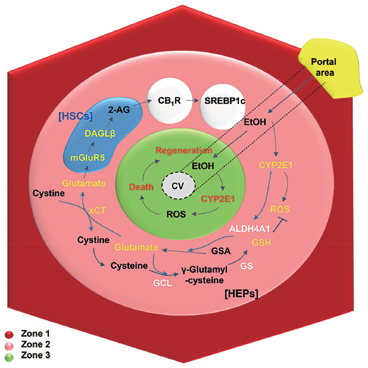
Chronic alcohol consumption induces the development of alcoholic steatosis in the liver, which is one of the most widespread liver diseases worldwide. During general alcohol metabolism, hepatocytes generate mitochondria- and cytochrome P450 2E1 (CYP2E1)-mediated reactive oxygen species (ROS) whose accumulation elicits activation of the hepatic anti-oxidant system, including glutathione (GSH). However, chronic alcohol consumption decreases GSH generation through cysteine deficiency by suppressing the methionine cycle and trans-sulfuration system, whereas it turns on an alternative defense pathway, such as the xCT transporter, to compensate for GSH shortage. The xCT transporter mediates the uptake of cystine coupled to the efflux of glutamate, leading to an increase in blood glutamate. In response to the elevated glutamate in the liver, the expression of metabotropic glutamate receptor 5 (mGluR5) is up-regulated in hepatic stellate cells (HSCs) along with enhanced production of 2-arachidonoylglycerol, which in turn stimulates cannabinoid receptor 1 (CB1R) on neighboring hepatocytes to increase de novo lipogenesis. On the other hand, blockade of mGluR5 and CB1R attenuates alcoholic steatosis. Interestingly, although the increased expression of CYP2E1-mediated xCT and ROS generation are mainly observed at the perivenous area (zone 3), fat accumulation is mostly detected at hepatic zone 2. To resolve this discrepancy, this review summarizes recent advances on glutamate/mGluR5-derived alcoholic steatosis and zone-dependently different responses to alcohol intake. In addition, the bidirectional loop pathway and its unique metabolic synapse between hepatocytes and HSCs are discussed.

Keywords: Alcoholic liver disease; Aldehyde dehydrogenase; Diacylglycerol lipase; Endocannabinoids; Homocysteine.


Conflict of interest statement

Conflicts of Interest

The authors have no conflicts to disclose.

Figures

Figure 1.
Figure 1.
Hepatic zonal-dependent fat accumulation in the early stage of ALD. On chronic ethanol consumption, ethanol is mainly metabolized in perivenous hepatocytes with high expression of CYP2E1, inducing metabolic stress-mediated death and regeneration of hepatocytes at zone 3. From zone 3 to 2, CYP2E1-mediated ROS can be eliminated by GSH generation through xCT-mediated uptake of cystine, while excreted glutamate stimulates mGluR5 of HSCs to produce 2-AG, followed by up-regulated lipogenesis of hepatocytes through CB1R-mediated SREBP1c expression. HSC, hepatic stellate cell; 2-AG, 2-arachidonoylglycerol; CB1R, cannabinoid receptor 1; SREBP1c, sterol regulatory element-binding protein 1; DAGL, diacylglycerol lipase; mGluR5, metabotropic glutamate receptor 5; CV, central vein; CYP2E1, cytochrome P450 2E1; ROS, reactive oxygen species; GSA, glutamic-γ-semialdehyde; GSH, glutathione; GS, glutathione synthase; GCL, glutamate cysteine ligase; HEPs, hepatocytes; ALD, alcoholic liver disease.
Figure 2.
Figure 2.
Hepatic zonal-dependent expression of CYP2E1 and ALDH4A1 in mice and patients with ALD. (A) After chronic alcohol consumption (4.5% of EtOH containing diet feeding for 8 weeks), fat accumulation is mainly observed at zone 2, whereas hepatic proliferation (Ki-67-positive) is mostly detected in zone 3 in stained liver sections of EtOH-fed mice. Arrows and asterisks indicate direction of blood flow and central veins, respectively. (B, C) Increased expression of CYP2E1 and ALDH4A1 is typically perceived around central veins of liver sections in EtOH-fed mice and alcoholic patients compared to their corresponding controls. Original magnification, ×200. H&E, Hematoxylin & Eosin; PT, portal area; CYP2E1, cytochrome P450 2E1; CV, central vein.

References

    1. Bajaj JS. Alcohol, liver disease and the gut microbiota. Nat Rev Gastroenterol Hepatol. 2019;16:235–246. - PubMed
    1. Seo W, Jeong WI. Hepatic non-parenchymal cells: master regulators of alcoholic liver disease? World J Gastroenterol. 2016;22:1348–1356. - PMC - PubMed
    1. Seitz HK, Bataller R, Cortez-Pinto H, Gao B, Gual A, Lackner C, et al. Alcoholic liver disease. Nat Rev Dis Primers. 2018;4:16. - PubMed
    1. Suh YG, Jeong WI. Hepatic stellate cells and innate immunity in alcoholic liver disease. World J Gastroenterol. 2011;17:2543–2551. - PMC - PubMed
    1. Spear BT, Jin L, Ramasamy S, Dobierzewska A. Transcriptional control in the mammalian liver: liver development, perinatal repression, and zonal gene regulation. Cell Mol Life Sci. 2006;63:2922–2938. - PMC - PubMed

LinkOut - more resources