. 2020 Oct 1;105(10):2368-2379.
doi: 10.3324/haematol.2019.239350.

The molecular basis for the prothrombotic state in sickle cell disease

Affiliations

The molecular basis for the prothrombotic state in sickle cell disease

Arun S Shet et al. Haematologica. .

Abstract

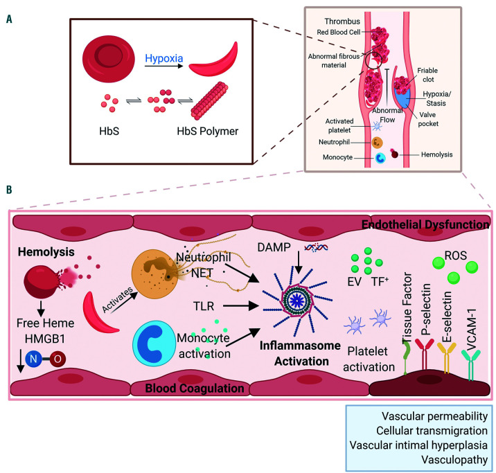
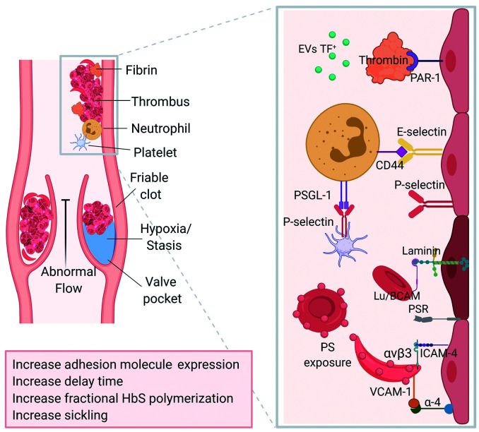
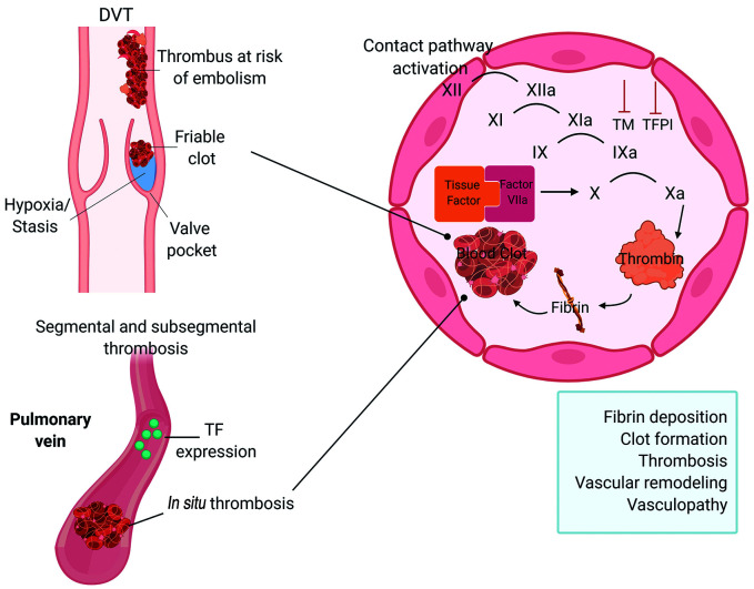
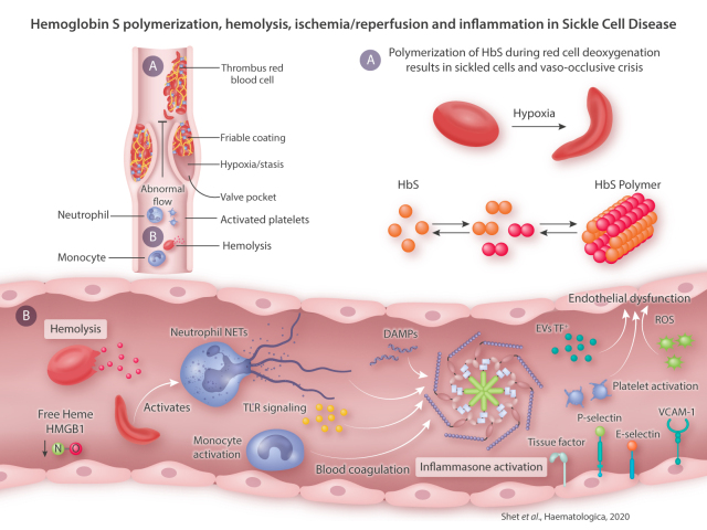
The genetic and molecular basis of sickle cell disease (SCD) has long since been characterized but the pathophysiological basis is not entirely defined. How a red cell hemolytic disorder initiates inflammation, endothelial dysfunction, coagulation activation and eventually leads to vascular thrombosis, is yet to be elucidated. Recent evidence has demonstrated a high frequency of unprovoked/recurrent venous thromboembolism (VTE) in SCD, with an increased risk of mortality among patients with a history of VTE. Here, we thoroughly review the molecular basis for the prothrombotic state in SCD, specifically highlighting emerging evidence for activation of overlapping inflammation and coagulation pathways, that predispose to venous thromboembolism. We share perspectives in managing venous thrombosis in SCD, highlighting innovative therapies with the potential to influence the clinical course of disease and reduce thrombotic risk, while maintaining an acceptable safety profile.


Figures

Figure 1.
Figure 1.
Sickle hemoglobin (HbS) polymerization, hemolysis and ischemia/reperfusion injury induce chronic inflammation. (A) Primary to sickle cell disease pathology is polymerization of HbS during red cell deoxygenation that results in sickled red cells and frequent painful vaso-occlusive crises. Hypoxia in the venous vasculature and valve pockets may worsen sickling mediated hemolysis and venous endothelial injury/inflammation. (B) Repeated sickling and unsickling episodes lead to intravascular hemolysis and release of free heme that consumes nitric oxide (NO). Sickle RBC also activate neutrophils among other cells, forming hetero and homotypic aggregates with blood cells that lead to vaso-occlusion in the post capillary venule. Endothelial damage and endothelial surface expression of adhesion and procoagulant molecules leads to transit delays increasing the potential for stasis and further sickling. Damage-associated molecular pattern (DAMP) molecules (free heme and high mobility group box 1 [HMGB1]) activate of various inflammatory pathways, e.g. NETosis, toll-like receptor (TLR) signaling, innate immune response and production of reactive oxidative species (ROS) lead to chronic inflammation. Repeated episodes of vaso-occlusive crisis (VOC) leads to ischemia followed by reperfusion mediating a well characterized injury response in the vascular endothelium. Figure created with BioRender.com. VCAM1: vascular cell adhesion molecule 1; EV TF: extracellular vesicle tissue factor; NET: neutrophil extracellular trap.
Figure 2.
Figure 2.
Inflammation and adhesion propagate stasis and contribute to fibrin deposition. Persistent inflammation leads to endothelial cell activation and endothelial dysfunction. Increased tissue factor (TF) and TF+ extracellular vesicle (EV) can generate thrombin and activate coagulation. Thrombin also activates cell surface protease activating receptors (PAR), worsening endothelial inflammation and dysfunction. Abnormal surface expression of adhesion molecules (P-selectin, E-selectin and V-CAM) and platelet activation facilitate heterotypic cellular interactions. Similarly, increased heterotypic and homotypic cellular adhesion interactions mediated via cell surface ligands E-selectin/CD44, Laminin/LuBCAM, α-4/VCAM-1, PSR/PS etc., promote vascular stasis and favor thrombosis. Overall this prothrombotic milieu favors red cell entrapment, vascular fibrin deposition, and thrombus formation. Figure created with BioRender.com. PSR: phosphatidylserine receptor; PAR1: protease activated receptor 1; PSGL-1: P-selectin glycoprotein ligand 1; EV TF: extracellular vesicle tissue factor; ICAM-4: intercellular adhesion molecule; Lu/BCAM: Lutheran/basal cell adhesion molecule; PS: phosphatidylserine.
Figure 3.
Figure 3.
The TF and contact pathways activate coagulation and generate thrombin. Abnormal expression of intravascular TF in sickle cell disease (SCD), the so called “blood borne TF”, triggers intravascular blood coagulation and pathological thrombosis. After binding with plasma factor VIIa, TF activates the extrinsic pathway of coagulation, generating thrombin, which mediates fibrin deposition, thrombosis and vascular remodeling. Besides lowered natural anticoagulant factor levels (thrombomodulin, protein C and S) also favor thrombosis. Activation of the contact pathway of coagulation by RBC phosphatidylserine (PS) exposure, cell-derived extracellular vesicles (EV), platelet polyphosphates, cfDNA and NET also sustain thrombin generation. TF and thrombin cause chronic inflammation possibly leading to endothelial injury, vascular permeability, angiogenesis and vascular remodeling, all of which are reflected by vasculopathy. Figure created with BioRender.com.
Graphical Abstract
Graphical Abstract

References

    1. Tisdale JF, Thein SL, Eaton WA. Treating sickle cell anemia. Science. 2020;367(6483):1198-1199. - PMC - PubMed
    1. Benz EJ, Mondoro TH, Gibbons GH. Accelerating the science of SCD therapies-is a cure possible? JAMA. 2019. Aug 8. [Epub ahead of print] - PubMed
    1. Collins FS. Curing HIV and sickle cell falls short if the most vulnerable populations are left out. Fortune. New York: Fortune Media IP Limited; 2020.
    1. Adams RJ, McKie VC, Hsu L, et al. Prevention of a first stroke by transfusions in children with sickle cell anemia and abnormal results on transcranial Doppler ultrasonography. N Engl J Med. 1998;339(1):5-11. - PubMed
    1. Ohene-Frempong K, Weiner SJ, Sleeper LA, et al. Cerebrovascular accidents in sickle cell disease: rates and risk factors. Blood. 1998;91(1):288-294. - PubMed

Publication types