

Cell-specific expression of Hfe determines the outcome of Salmonella enterica serovar Typhimurium infection in mice
- PMID: 33054105
- PMCID: PMC8634192
- DOI: 10.3324/haematol.2019.241745
Cell-specific expression of Hfe determines the outcome of Salmonella enterica serovar Typhimurium infection in mice
Abstract
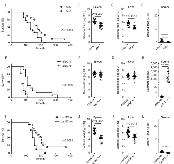
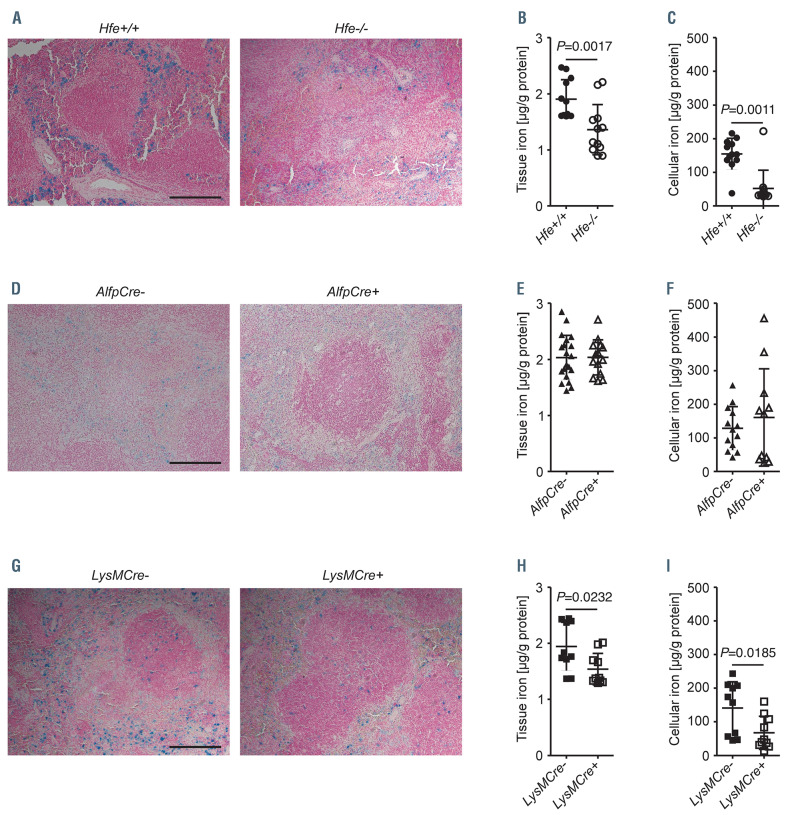
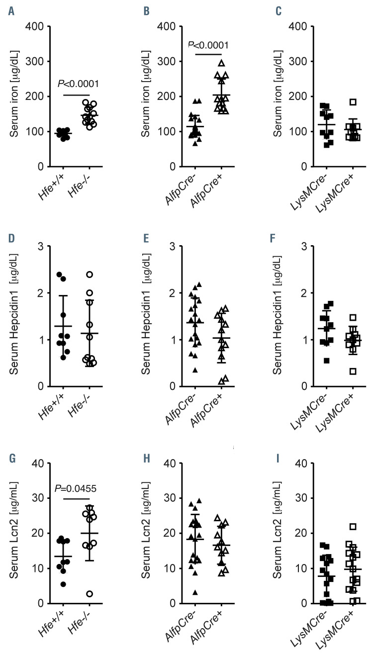
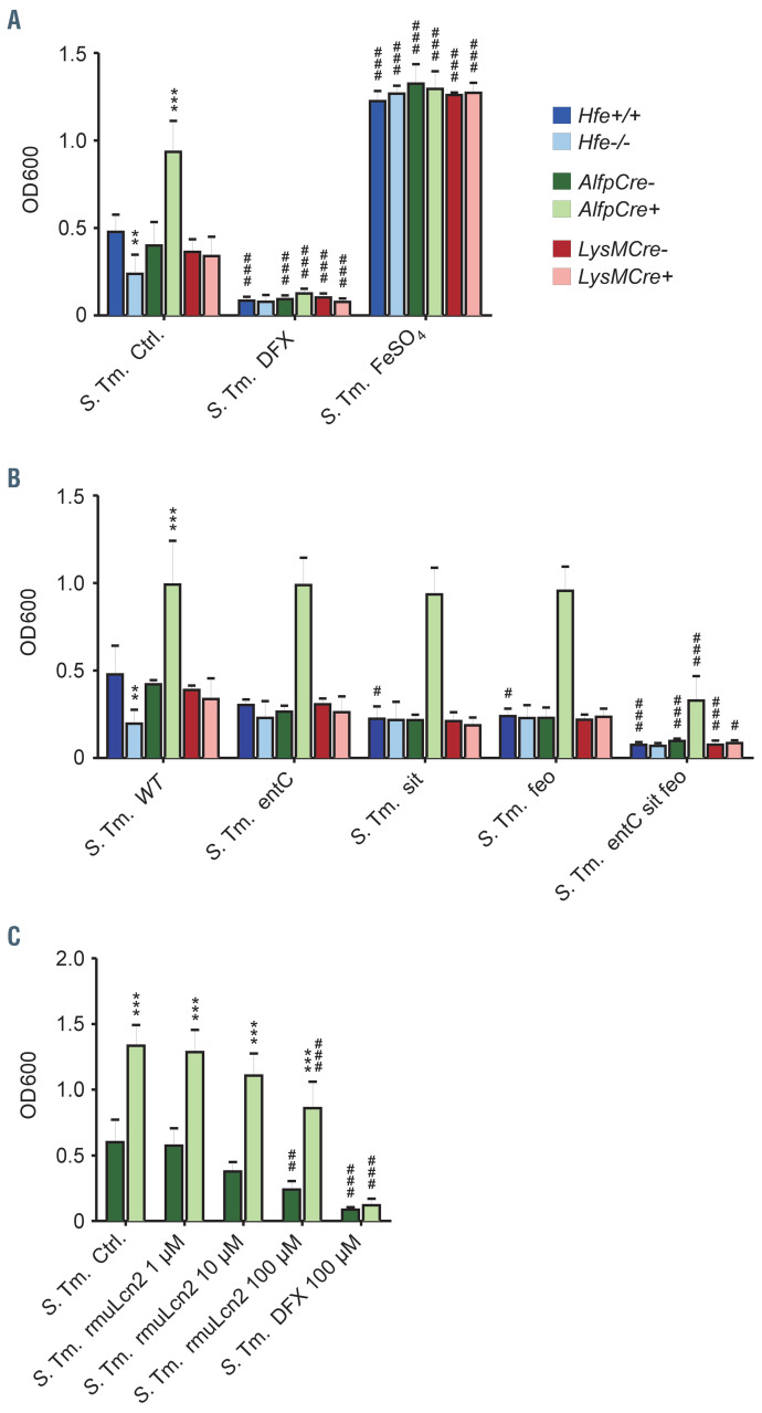
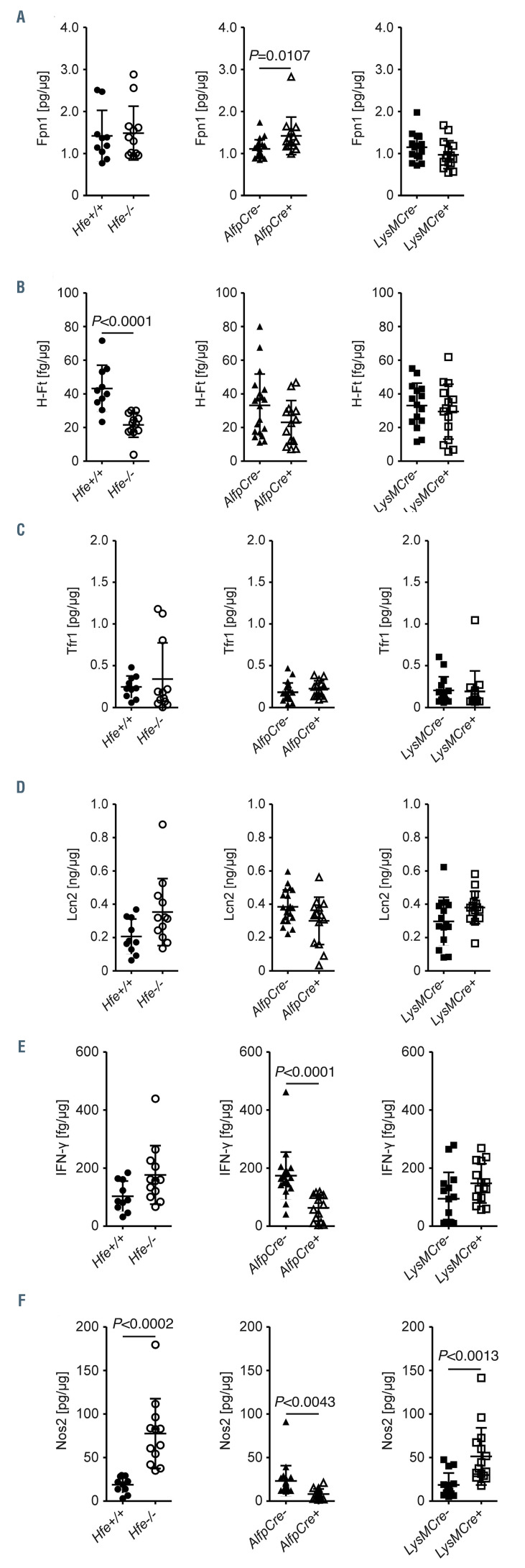
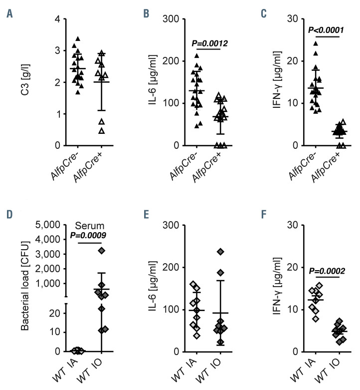
Mutations in HFE cause hereditary hemochromatosis type I hallmarked by increased iron absorption, iron accumulation in hepatocytes and iron deficiency in myeloid cells. HFE encodes an MHC-I like molecule, but its function in immune responses to infection remains incompletely understood. Here, we investigated putative roles of Hfe in myeloid cells and hepatocytes, separately, upon infection with Salmonella Typhimurium, an intracellular bacterium with iron-dependent virulence. We found that constitutive and macrophage-specific deletion of Hfe protected infected mice. The propagation of Salmonella in macrophages was reduced due to limited intramacrophage iron availability for bacterial growth and increased expression of the anti-microbial enzyme nitric oxide synthase-2. By contrast, mice with hepatocyte-specific deletion of Hfe succumbed earlier to Salmonella infection because of unrestricted extracellular bacterial replication associated with high iron availability in the serum and impaired expression of essential host defense molecules such as interleukin-6, interferon-γ and nitric oxide synthase-2. Wild-type mice subjected to dietary iron overload phenocopied hepatocyte-specific Hfe deficiency suggesting that increased iron availability in the serum is deleterious in Salmonella infection and underlies impaired host immune responses. Moreover, the macrophage-specific effect is dominant over hepatocyte-specific Hfe-depletion, as Hfe knock-out mice have increased survival despite the higher parenchymal iron load associated with systemic loss of Hfe. We conclude that cell-specific expression of Hfe in hepatocytes and macrophages differentially affects the course of infections with specific pathogens by determining bacterial iron access and the efficacy of anti-microbial immune effector pathways. This may explain the high frequency and evolutionary conservation of human HFE mutations.
Figures
Similar articles
-
Genetic and Dietary Iron Overload Differentially Affect the Course of Salmonella Typhimurium Infection.Front Cell Infect Microbiol. 2017 Apr 11;7:110. doi: 10.3389/fcimb.2017.00110. eCollection 2017. Front Cell Infect Microbiol. 2017. PMID: 28443246 Free PMC article.
-
Absence of functional Hfe protects mice from invasive Salmonella enterica serovar Typhimurium infection via induction of lipocalin-2.Blood. 2009 Oct 22;114(17):3642-51. doi: 10.1182/blood-2009-05-223354. Epub 2009 Aug 21. Blood. 2009. PMID: 19700664 Free PMC article.
-
Genetic disruption of NRF2 promotes the development of necroinflammation and liver fibrosis in a mouse model of HFE-hereditary hemochromatosis.Redox Biol. 2017 Apr;11:157-169. doi: 10.1016/j.redox.2016.11.013. Epub 2016 Dec 1. Redox Biol. 2017. PMID: 27936457 Free PMC article.
-
Pathophysiological consequences and benefits of HFE mutations: 20 years of research.Haematologica. 2017 May;102(5):809-817. doi: 10.3324/haematol.2016.160432. Epub 2017 Mar 9. Haematologica. 2017. PMID: 28280078 Free PMC article. Review.
-
HFE gene: Structure, function, mutations, and associated iron abnormalities.Gene. 2015 Dec 15;574(2):179-92. doi: 10.1016/j.gene.2015.10.009. Epub 2015 Oct 9. Gene. 2015. PMID: 26456104 Free PMC article. Review.
Cited by
-
Regulation of Th1 T Cell Differentiation by Iron via Upregulation of T Cell Immunoglobulin and Mucin Containing Protein-3 (TIM-3).Front Immunol. 2021 May 24;12:637809. doi: 10.3389/fimmu.2021.637809. eCollection 2021. Front Immunol. 2021. PMID: 34108960 Free PMC article.
-
Iron and the immune system.Nat Rev Immunol. 2025 Jun 16. doi: 10.1038/s41577-025-01193-y. Online ahead of print. Nat Rev Immunol. 2025. PMID: 40524018 Review.
-
DMT1 Protects Macrophages from Salmonella Infection by Controlling Cellular Iron Turnover and Lipocalin 2 Expression.Int J Mol Sci. 2022 Jun 17;23(12):6789. doi: 10.3390/ijms23126789. Int J Mol Sci. 2022. PMID: 35743233 Free PMC article.
-
Iron Metabolism in Aging and Age-Related Diseases.Int J Mol Sci. 2022 Mar 25;23(7):3612. doi: 10.3390/ijms23073612. Int J Mol Sci. 2022. PMID: 35408967 Free PMC article. Review.
References
-
- Pietrangelo A. Hereditary hemochromatosis-- a new look at an old disease. N Engl J Med. 2004;350(23):2383-2397. - PubMed
-
- Weiss G. Genetic mechanisms and modifying factors in hereditary hemochromatosis. Nat Rev Gastroenterol Hepatol.; 7(1):50-58. - PubMed
-
- Cairo G, Recalcati S, Montosi G, Castrusini E, Conte D, Pietrangelo A. Inappropriately high iron regulatory protein activity in monocytes of patients with genetic hemochromatosis. Blood. 1997;89(7):2546-2553. - PubMed
-
- Montosi G, Paglia P, Garuti C, et al. . Wildtype HFE protein normalizes transferrin iron accumulation in macrophages from subjects with hereditary hemochromatosis. Blood. 2000;96(3):1125-1129. - PubMed
MeSH terms
Substances
Grants and funding
LinkOut - more resources
Full Text Sources
Medical
Research Materials
