. 2021 Nov 1;106(11):2859-2873.
doi: 10.3324/haematol.2020.256347.

Characterization and evolutionary origin of novel C2H2 zinc finger protein (ZNF648) required for both erythroid and megakaryocyte differentiation in humans

Affiliations

Characterization and evolutionary origin of novel C2H2 zinc finger protein (ZNF648) required for both erythroid and megakaryocyte differentiation in humans

Daniel C J Ferguson et al. Haematologica. .

Abstract

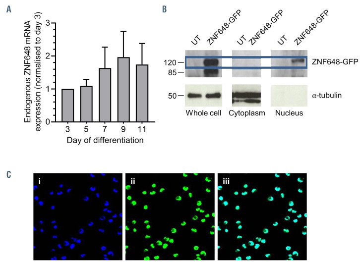
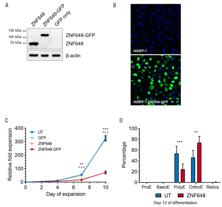
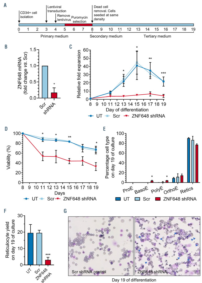
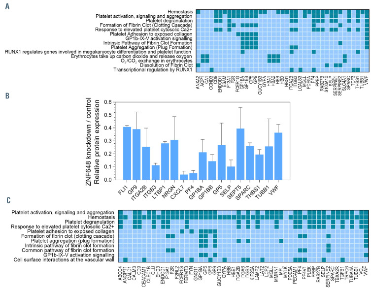
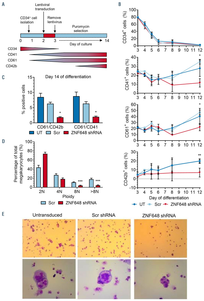
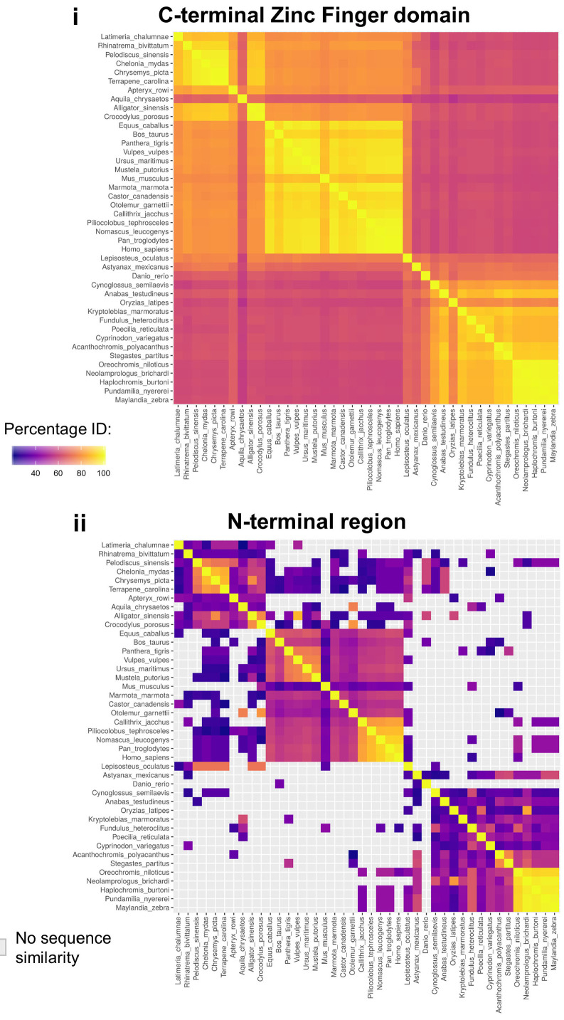
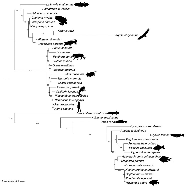
Human ZNF648 is a novel poly C-terminal C2H2 zinc finger protein identified amongst the most dysregulated proteins in erythroid cells differentiated from iPSC. Its nuclear localisation and structure indicate it is likely a DNA-binding protein. Using a combination of ZNF648 overexpression in an iPSC line and primary adult erythroid cells, ZNF648 knockdown in primary adult erythroid cells and megakaryocytes, comparative proteomics and transcriptomics we show that ZNF648 is required for both erythroid and megakaryocyte differentiation. Orthologues of ZNF648 were detected across Mammals, Reptilia, Actinopterygii, in some Aves, Amphibia and Coelacanthiformes suggesting the gene originated in the common ancestor of Osteichthyes (Euteleostomi or bony fish). Conservation of the C-terminal zinc finger domain is higher, with some variation in zinc finger number but a core of at least six zinc fingers conserved across all groups, with the N-terminus recognisably similar within but not between major lineages. This suggests the N-terminus of ZNF648 evolves faster than the C-terminus, however this is not due to exon-shuffling as the entire coding region of ZNF648 is within a single exon. As for other such transcription factors, the N-terminus likely carries out regulatory functions, but showed no sequence similarity to any known domains. The greater functional constraint on the zinc finger domain suggests ZNF648 binds at least some similar regions of DNA in the different organisms. However, divergence of the N-terminal region may enable differential expression, allowing adaptation of function in the different organisms.


Figures

Figure 1.
Figure 1.
Expression and localisation of ZNF648 during erythroid differentiation. (A) Expression of ZNF648 transcript levels at days 3, 5, 7, 9 and 11 in culture analysed by quantitative polymerase chain reaction (qPCR), n=2. (B) Western blot of whole cell lysate, cytoplasmic fraction, and nuclear fraction of control K562 cells and K562 cells transduced with ZNF648-GFP probed with antibody to green fluorescent protein (GFP). Antibody to -tubulin was used as a control for cytoplasmic fraction protein loading. Molecular weight (MW) markers shown on left hand side. (C) images of K562 cells transduced with ZNF648-GFP (i) DAPI staining (ii) ZNF648-GFP fluorescence (iii) Merged image of panels i and ii. Images obtained using a Leica SP5 confocal microscope. Magnification 400x.
Figure 2.
Figure 2.
Exogenous expression of ZNF648 in the induced pluripotent stem cells line HiDEP-1 impedes proliferation and advances differentiation. HiDEP-1 cells were transduced with ZNF648, ZNF648-GFP or a control green fluorescent protein (GFP) construct. (A) Western blot of HiDEP-1 cells probed with ZNF648 antibody (Abcam). Molecular wieght (MW) markers shown on left hand side (B) Images of control and HiDEP-1 cells transduced with ZNF648-GFP; DAPI nuclear staining in blue. (C) Relative fold expansion of ZNF648 and ZNF648-GFP expressing HiDEP-1 cells compared to untransduced (UT) and GFP control cells. *GFP vs. ZNF648, +GFP vs. ZNF648-GFP. **/++ P<0.01; ***/+++ P<0.001, n=2 each for ZNF648 and ZNF648-GFP ± standard deviation, t-test. (D) proportion of HiDEP-1 cells at each stage of differentiation present at day 12 of culture. ProE: proerythroblast; BasoE: basophilic erythroblast; PolyE: polychromatic erythroblast; OrthoE: orthochromatic erythroblast; Retics: reticulocytes. **P<0.01, ***P<0.001, 2-way ANOVA. N=3 ± standard deviation.
Figure 3.
Figure 3.
ZNF648 knockdown impedes erythroid differentiation. Erythroblasts differentiated from adult peripheral blood (PB) CD34+ cells at day 3 in culture were transduced with scrambled (Scr) short hairpin RNA (shRNA) as control or ZNF648 shRNA, puromycin selected and seeded at same cell density along with untransduced cells (UT) that served as a further control. (A) Schematic of experimental design. (B) quantitative polymerase chain reaction (qPCR) of ZNF648 transcript levels normalised to Scr control. (C) Relative fold expansion during differentiation compared to cell number at day 8 in culture. (D) Viability of cells during differentiation assessed by trypan blue exclusion. (E) Proportion of cells at different stages of differentiation at day 19 in culture. ProE: proerythroblast; BasoE: basophilic erythroblast; PolyE: polychromatic erythroblast; OrthoE: orthochromatic erythroblast; Retics: reticulocytes. (F) Reticulocyte yield at day 19 of culture. (G) Morphology of cells at day 19 of culture stained with May- Grünwald-Giemsa, representative images of 3 independent cultures. *P<0.05, **P<0.01, ***P<0.001, ttest, n=3 ± standard devation.
Figure 4.
Figure 4.
Megakaryocyte-associated proteins and pathway classification of transcripts and proteins decreased in level following ZNF648 knockdown. Total RNA and whole cell protein lysates were prepared from erythroid cells differentiated from adult peripheral blood (PB) CD34+ cells transduced with scrambled (Scr) or ZNF648 short hairpin RNA (shRNA), at day 8 in culture. RNA was used to screen human genome arrays, with transcripts decreased in level by ≥2-fold following ZNF648 knockdown (KD) entered into Reactome. Data from three independent KD experiments (A). Tryptic peptides were prepared from cell lysates and labeled with Tandem Mass Tags followed by nanoscale liquid chromatography coupled to tandem mass spectrometry (nano-LC MS/MS) with (B) relative decrease in level of megakaryocyte associated proteins following ZNF648 KD, identified amongst the 123 unique proteins decreased by ≥2-fold. Data from two independent KD experiments ± standard deviation, and (C) Reactome analysis of the proteins decreased in level. In (A) and (C) dark shades indicate genes/proteins apportioned to the pathway shown on the left. Pathways shown with minimum probability of over representation in the dataset, corrected for false discovery rate of P<0.006 for transcripts and P<1.42x10⁻⁵ for proteins.
Figure 5.
Figure 5.
Pathway classification of proteins increased in level following ZNF648 knockdown. Tandem Mass Tag labeling and nanoscale liquid chromatography coupled to tandem mass spectrometry (nano-LC MS/MS) was performed on whole cell lysates from erythroid cells transduced with ZNF648 or scrambled (Scr) (control) short hairpin RNA (shRNA) at day 8 in culture. Proteins increased in level following ZNF648 knockdown were entered into Reactome. Dark red indicates proteins apportioned to the pathway shown on the left. Top 20 pathways shown with minimum probability of over representation in the dataset, corrected for false discovery rate, of P<2.11x10⁻⁵.
Figure 6.
Figure 6.
ZNF648 knockdown impedes megakaryocyte differentiation. Isolated adult peripheral blood CD34+ cells were transferred to megakaryocyte differentiation medium and transduced with scrambled (Scr) short hairpin RNA (shRNA) as control or ZNF648 shRNA followed by puromycin selection. Untransduced cells served as a further control. (A) Schematic of experimental design and expression profile of CD34, CD41, CD61 and CD42b during normal megakaryopoiesis, (B) expression of membrane markers CD34, CD41 (platelet glycoprotein IIb), CD61 (platelet glycoproteins IIIa) and CD42b (GPIb ) analysed by flow cytometry with antibodies CD41- PE, CD61-APC (both from Biolegend), CD34-VB BD and CD42b-PE BD (both from Pharming). Data was acquired with a MacsQuant VYB Analyser using a plate reader, n=6. (C) Proportion of CD41/CD61 and CD61/CD42b positive cells on day 14 of culture, n=6 ± standard error of the mean. (D) Ploidy status of cells at day 14 of culture, n=3. (E) Morphology of cells at day 14 of culture stained with May-Grünwald-Giemsa. Upper panel 100x magnification, lower panel 400x magnification. Images representative of three independent cultures. *P<0.05, **P<0.01, ***P<0.001, Student’s t-test.
Figure 7.
Figure 7.
Pairwise sequence identity and phylogeny of representative ZNF648 orthologues across Osteichthyes. (A) (i) C-terminus (human residues 279-568;), (ii) N-terminus (human residues 1-278). Warm colors indicate higher pairwise percentage identity. The C-terminus (containing the ZnF motifs) is conserved across Osteichthyes, but the N-terminus is much less conserved, showing high identity only within major lineages.
Figure 8.
Figure 8.
ZNF648 phylogenetic tree. A maximum likelihood phylogeny of ZNF648 was inferred (949 aligned sites/1134 positions/42 taxa) using the best-fitting LG+C60+G+F model in IQ-Tree 1.6.10. Branch supports are 1,000 ultrafast bootstraps. ZNF648 is conserved across the major lineages of Osteichthyes (Euteleostomi), but several lineage-specific losses have occurred. The topology of the tree is congruent with our understanding of species relationships, suggesting that the gene traces back to the common ancestor of Osteichthyes.

References

    1. Iuchi S. Three classes of C2H2 zinc finger proteins. Cell Mol Life Sci. 2001;58(4):625-635. - PMC - PubMed
    1. Klug A. The discovery of zinc fingers and their development for practical applications in gene regulation and genome manipulation. Q Rev Biophys. 2010;43(1):1-21. - PubMed
    1. Vaquerizas JM, Kummerfeld SK, Teichmann SA, et al. . A census of human transcription factors: function, expression and evolution. Nat Rev Genet. 2009;10(4):252-263. - PubMed
    1. Kim SI, Bresnick EH. Transcriptional control of erythropoiesis: emerging mechanisms and principles. Oncogene. 2007;26(47):6777-6794. - PubMed
    1. Siatecka M, Bieker JJ. The multifunctional role of EKLF/KLF1 during erythropoiesis. Blood. 2011;118(8):2044-2054. - PMC - PubMed

Publication types