. 2021 Jan 1;20(1):704-714.
doi: 10.1021/acs.jproteome.0c00614. Epub 2020 Oct 15.

Temporal Proteomic Profiling of SH-SY5Y Differentiation with Retinoic Acid Using FAIMS and Real-Time Searching

Affiliations

Temporal Proteomic Profiling of SH-SY5Y Differentiation with Retinoic Acid Using FAIMS and Real-Time Searching

Tian Zhang et al. J Proteome Res. .

Abstract

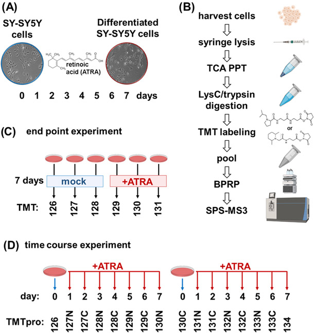
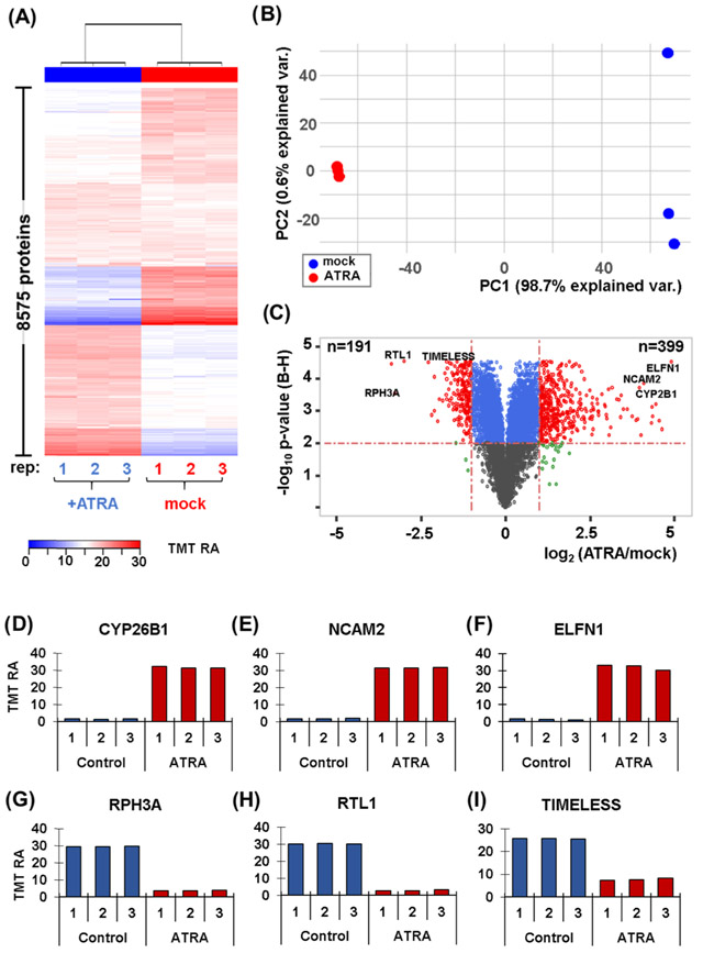
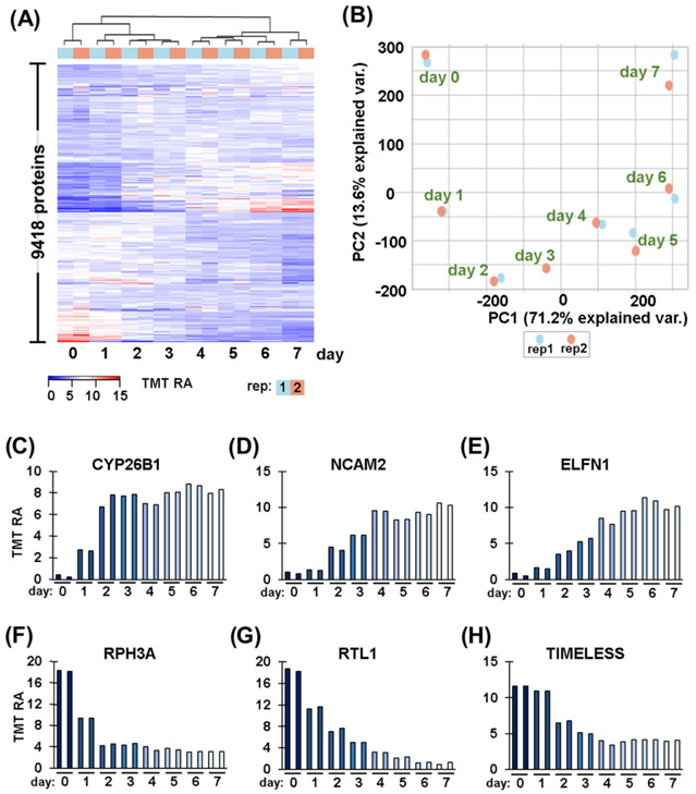
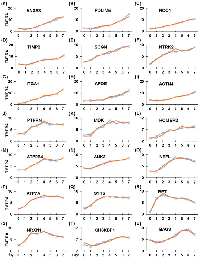
The SH-SY5Y cell line is often used as a surrogate for neurons in cell-based studies. This cell line is frequently differentiated with all-trans retinoic acid (ATRA) over a 7-day period, which confers neuron-like properties to the cells. However, no analysis of proteome remodeling has followed the progress of this transition. Here, we quantitatively profiled over 9400 proteins across a 7-day treatment with retinoic acid using state-of-the-art mass spectrometry-based proteomics technologies, including FAIMS, real-time database searching, and TMTpro16 sample multiplexing. Gene ontology analysis revealed that categories with the highest increases in protein abundance were related to the plasma membrane/extracellular space. To showcase our data set, we surveyed the protein abundance profiles linked to neurofilament bundle assembly, neuron projections, and neuronal cell body formation. These proteins exhibited increases in abundance level, yet we observed multiple patterns among the queried proteins. The data presented represent a rich resource for investigating temporal protein abundance changes in SH-SY5Y cells differentiated with retinoic acid. Moreover, the sample preparation and data acquisition strategies used here can be readily applied to any analogous cell line differentiation analysis.

Keywords: ATRA; SPS-MS3; TMTpro; eclipse; multinotch; retinoic acid.


Figures

Figure 1.
Figure 1.
Experimental overview. (A) SH-SY5Y has been shown to differentiate when treated with all-trans-retinoic acid (ATRA) over a 7-day period and to acquire neuron-like properties. (B) Experimental workflow. Samples were processed using the SL-TMT protocol (45). (C) Layout of TMT6-plex end-point experiment. Triplicates of untreated and 7-day retinoic acid-treated SH-SY5Y cells were arranged in a 3 × 2 TMT6-plex experiment. (D) Layout of TMTpro16-plex time-course. SH-SY5Y cells treated with retinoic acid were harvested over the course of 7 days. Biological replicate samples from Day 0 to Day 7 were arranged in a TMTpro16-plex. ATRA, all-trans retinoic acid; BPRP, basic pH reversed-phase; TCA PPT, trichloroacetic acid precipitation.
Figure 2.
Figure 2.
Proteome profiling of SH-SY5Y cells with and without a 7-day retinoic acid treatment. (A) Heat map illustrates the relative abundance of the 8,575 proteins quantified in this experiment. Each protein abundance value sums to 100 across the six channels. (B) Principal components analysis (PCA) of the six samples depicts the variance explained by the first two principal components (PC). (C) Volcano plot illustrating the Benjamini–Hochberg (BH)-corrected p-value of retinoic acid (ATRA) vs. mock control and the fold change for these values. Highlighted on the volcano plot are six of the most altered proteins resulting from retinoic acid treatment. These proteins include: (D) CYP26B1, cytochrome P450 26B1; (E) NCAM2, neural cell adhesion molecule 2; (F) ELFN1, extracellular leucine-rich repeat and fibronectin type III.(G) RPH3A, rabphilin-3A; (H) RTL1, retrotransposon-like protein 1; and (I) TIMELESS.
Figure 3.
Figure 3.
Temporal differences in protein expression. (A) Heat map illustrates the relative abundance of the 9418 proteins quantified in this time-course experiment. The abundance values for each protein sum to 100 across the 16 channels. (B) Principal components analysis (PCA) of the 16 samples depicts the variance explained by the first two principal components (PC). Temporal expression patterns of proteins highlighted in Figure 2: (C) CYP26B1, cytochrome P450 26B1; (D) NCAM2, neural cell adhesion molecule 2; (E) ELFN1, extracellular leucine-rich repeat and fibronectin type III, (F) RPH3A, rabphilin-3A; (G) RTL1, retrotransposon-like protein 1; and (H) TIMELESS.
Figure 4.
Figure 4.
Temporal protein profiles of neuron-related proteins. (A–U) Proteins associated with neurons, particularly neurite outgrowth, are highlighted. These proteins include: (A) Annexin A3, ANXA3; (B) PDZ and LIM domain protein 5, PDLIM5; (C) NAD(P)H dehydrogenase [quinone] 1, NQO1; (D) Metalloproteinase inhibitor 2, TIMP2; (E) Secretagogin, SCGN; (F) BDNF/NT-3 growth factors receptor, NTRK2; (G) Integrin alpha1, ITGA1; (H) Apolipoprotein E, APOE; (I) α-actinin-4, ACTN4; (J) Receptor-type tyrosine-protein phosphatase-like N, PTPRN; (K) Midkine, MDK; (L) Homer protein, HOMER2; (M) Plasma membrane calcium-transporting ATPase 4, ATP2B4; (N) Ankyrin-3, ANK3; (O) Neurofilament light polypeptide, NEFL; (P) Copper-transporting ATPase 1, ATP7A; (Q) Synaptotagmin-5, SYT5; (R) Proto-oncogene tyrosine-protein kinase receptor, RET; (S) Neurexin-1, NRXN1; (T) SH3 domain-containing kinase-binding protein 1, SH3KBP1; and (U) BAG family molecular chaperone regulator 3, BAG3.

References

    1. Zhang M; Zhang YQ; Wei XZ; Lee C; Huo DS; Wang H; Zhao ZY Differentially expressed long-chain noncoding RNAs in human neuroblastoma cell line (SH-SY5Y): Alzheimer’s disease cell model. J. Toxicol. Environ. Health, Part A 2019, 82, 1052–1060. - PubMed
    1. de Medeiros LM; De Bastiani MA; Rico EP; Schonhofen P; Pfaffenseller B; Wollenhaupt-Aguiar B; Grun L; Barbe-Tuana F; Zimmer ER; Castro MAA; Parsons RB; Klamt F Cholinergic Differentiation of Human Neuroblastoma SH-SY5Y Cell Line and Its Potential Use as an In vitro Model for Alzheimer’s Disease Studies. Mol. Neurobiol 2019, 56, 7355–7367. - PubMed
    1. Azzolin VF; Barbisan F; Lenz LS; Teixeira CF; Fortuna M; Duarte T; Duarte M; da Cruz IBM Effects of Pyridostigmine bromide on SH-SY5Y cells: An in vitro neuroblastoma neurotoxicity model. Mutat. Res., Genet. Toxicol. Environ. Mutagen 2017, 823, 1–10. - PubMed
    1. Xie HR; Hu LS; Li GY SH-SY5Y human neuroblastoma cell line: in vitro cell model of dopaminergic neurons in Parkinson’s disease. Chin. Med. J 2010, 123, 1086–1092. - PubMed
    1. Kovalevich J; Langford D Considerations for the Use of SH-SY5Y Neuroblastoma Cells in Neurobiology. Methods in Molecular Biology; Humana Press: Totowa, NJ, 2013; Vol. 1078, pp 9–21. - PMC - PubMed

Publication types