. 2020 Oct 28;7(5):ENEURO.0423-19.2020.
doi: 10.1523/ENEURO.0423-19.2020. Print 2020 Sep/Oct.

Chemogenetic Inhibition of Infralimbic Prefrontal Cortex GABAergic Parvalbumin Interneurons Attenuates the Impact of Chronic Stress in Male Mice

Affiliations

Chemogenetic Inhibition of Infralimbic Prefrontal Cortex GABAergic Parvalbumin Interneurons Attenuates the Impact of Chronic Stress in Male Mice

Nawshaba Nawreen et al. eNeuro. .

Abstract

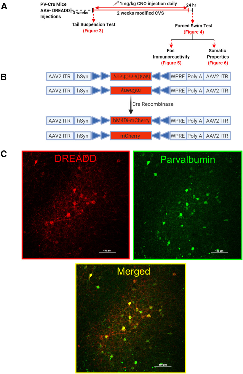
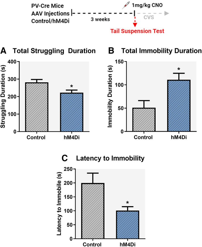
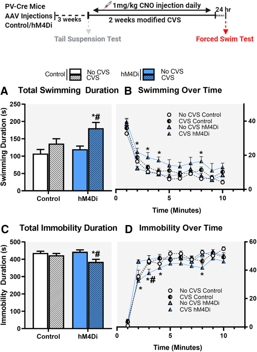
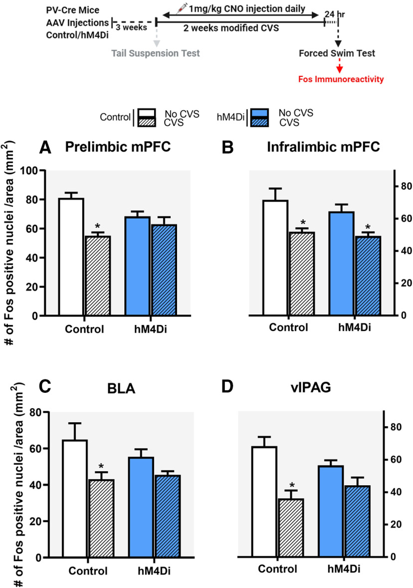
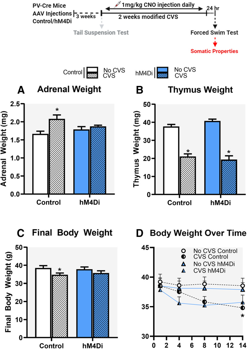
Hypofunction of the prefrontal cortex (PFC) contributes to stress-related neuropsychiatric illnesses. Mechanisms leading to prefrontal hypoactivity remain to be determined. Prior evidence suggests that chronic stress leads to an increase in activity of parvalbumin (PV) expressing GABAergic interneurons (INs) in the PFC. The purpose of the study was to determine whether reducing PV IN activity in the Infralimbic (IL) PFC would prevent stress-related phenotypes. We used a chemogenetic approach to inhibit IL PFC PV INs during stress. Mice were first tested in the tail suspension test (TST) to determine the impact of PV IN inhibition on behavioral responses to acute stress. The long-term impact of PV IN inhibition during a modified chronic variable stress (CVS) was tested in the forced swim test (FST). Acute PV IN inhibition reduced active (struggling) and increased passive coping behaviors (immobility) in the TST. In contrast, inhibition of PV INs during CVS increased active and reduced passive coping behaviors in the FST. Moreover, chronic inhibition of PV INs attenuated CVS-induced changes in Fos expression in the prelimbic cortex (PrL), basolateral amygdala (BLA), and ventrolateral periaqueductal gray (vlPAG) and also attenuated adrenal hypertrophy and body weight loss associated with chronic stress. Our results suggest differential roles of PV INs in acute versus chronic stress, indicative of distinct biological mechanisms underlying acute versus chronic stress responses. Our results also indicate a role for PV INs in driving chronic stress adaptation and support literature evidence suggesting cortical GABAergic INs as a therapeutic target in stress-related illnesses.

Keywords: DREADDs; GABA; interneurons; parvalbumin; prefrontal cortex; stress coping.


Figures

Figure 1.
Figure 1.
Experimental design and targeting of PV INs in the PFC using DREADDs. A, Experimental design and timeline. C57BL/6J PV-Cre mice ∼7.5 months of age underwent surgery to inject AAV2-hM4Di-mCherry (inhibitory DREADD) or AAV2-mCherry (control virus). Mice were allowed three weeks to recover to enable sufficient time for DREADD expression. Animals were then subjected to CVS procedure twice a day for 14 d or served as controls. The first stressor was a TST to determine acute effects of PV IN inhibition in animals within the CVS group. Animals were dosed with 1 mg/kg CNO before each stressor to inhibit PV INs during the CVS procedure; 24 h after the end of CVS, animals were subjected to FST, following which mice were euthanized, body was perfused, and brains and organs were collected. No CNO was administered during the testing phase in FST. B, Design of AAV2-hSyn-hM4Di-mCherry (top) and AAV2-hSyn-mCherry (bottom) vectors employing the DIO strategy. Two pairs of heterotypic, antiparallel loxP recombination sites (blue triangles) achieve Cre-mediated transgenes inversion and expression under the control of hSyn promoter. ITR, left-inverted terminal repeat; hSyn, human synapsin; WPRE, woodchuck hepatitis DREADD posttranscriptional regulatory element. C, Successful Cre-mediated recombination of DREADDs demonstrated by presence of red mCherry (left); green fluorescence identifies PV INs (right); hM4Di receptors selectively expressed in PV INs as illustrated by mCherry (red) and PV (green) co-expression (merged, yellow, bottom middle image). Scale bar: 100 μm.
Figure 2.
Figure 2.
Injection site, viral spread. and targeting of IL. A, Injection site and viral spread mapped onto respective mouse bregma coordinates, following bilateral injection of hM4Di DREADD into the IL of PV-Cre mice (n = 9). Light green rectangular box depicts the IL region with the injection sites restricted to the region. Each animal’s injection site and viral spread has been represented by a unique color. B, Representative image from one animal demonstrating the viral spread in PFC detected by red mCherry fluorescence. Image shows the spread was restricted to the IL with the white dashed lines outlining the IL cortex region of the PFC. Scale bars: 1000 and 500 μm. C, Percentage of DREADD transfected PV INs in the IL of PV-Cre mice. The percentage of infected cells in IL was calculated by dividing the number of mCherry-positive cells in IL by the total number of mCherry-positive cells in a specific focal plane and bregma coordinate. Brain bregma coordinates used for IL cytoarchitecture were taken from Franklin and Paxinos (2007). *indicates significant effect p < 0.05 compared to outside IL group. Values represent mean ± SEM, n = 9 per group.
Figure 3.
Figure 3.
Impact of acute chemogenetic inhibition of PV IN in the IL mPFC on coping behavior in the TST. Acute inhibition of PV INs in the IL mPFC during TST, reduced total time spent struggling (A), increased total time spent immobile (B), and reduced latency to immobility (C). All mice were treated with CNO (1 mg/kg, i.p.) 30 min before TST. Behaviors were analyzed for a total time of 6 min. Values represent mean ± SEM, n = 9–10 per group; * indicates significant effect p < 0.05 versus corresponding control group.
Figure 4.
Figure 4.
Effects of chronic chemogenetic inhibition of PV INs during CVS on coping behavior in the FST following CVS. Chronic inhibition of PV INs in the IL mPFC during CVS, resulted in increased total time spent swimming (A) and decreased total time spent immobile (C) in the FST. B, D, Changes in swimming and immobility behavior, respectively, over the 10 min of FST. Values represent mean ± SEM, n = 9–10 per group; * indicates planned comparison significant effect p < 0.05 versus corresponding No CVS hM4Di group; # indicates planned comparison significant effect p < 0.05 versus corresponding CVS Control group. Extended Data Figure 4-1 demonstrates chronic inhibition of PV INs did not affect locomotor activity. Extended Data Figure 4-2 demonstrates chronic CNO administration did not lead to any changes in FST behavior following CVS or control. Extended Data Figure 4-3 demonstrates CVS effects on FST did not depend on the age of the animal and also shows chronic injection stress and housing conditions had no effect on FST behavior in control animals.
Figure 5.
Figure 5.
Fos immunoreactivity in the PrL, IL, BLA, and vlPAG. Chronic inhibition of PV INs in the IL mPFC during CVS, attenuated CVS-mediated reduction in Fos expression in the PrL, BLA, and vlPAG, respectively (A, C, D) but did not prevent CVS-mediated reduction in Fos expression in the IL cortex (B) following FST. Values are presented as mean ± SEM; n = 7–10 per group; * indicates significant result p < 0.05 post hoc (A) and planned comparisons (C, D) compared with respective No CVS Control groups. PrL, IL, BLA, and vlPAG stands for prelimbic, infralimbic, basolateral amygdala and ventrolateral periaqueductal grey, respectively. Extended Data Figure 5-1 demonstrates Fos expression in various other brain regions where no significant effect of PV IN modulation was observed.
Figure 6.
Figure 6.
Impact of chronic stress on organ and body weights. Chronic inhibition of PV INs in the IL mPFC during CVS, attenuated CVS mediated increases in adrenal gland weight (A) and attenuated CVS mediated decreases in body weight (C, D). No change in CVS-induced decreases in thymus weight was observed following PV IN inhibition (B). Data are presented as absolute organ and body weights. Values represent mean ± SEM; n = 9–10 per group; * indicates planned comparisons significant effect p < 0.05 versus corresponding No CVS Control groups.

References

    1. Baldessarini RJ, Centorrino F, Huston Lyons D, Cohen BM, Flood JG, Volpicelli SA (1993) Tissue concentrations of clozapine and its metabolites in the rat. Neuropsychopharmacology 9:117–124. 10.1038/npp.1993.50 - DOI - PubMed
    1. Berg L, Eckardt J, Masseck OA (2019) Enhanced activity of pyramidal neurons in the infralimbic cortex drives anxiety behavior. PLoS One 14:e0210949. 10.1371/journal.pone.0210949 - DOI - PMC - PubMed
    1. Berton O, Covington HE, Ebner K, Tsankova NM, Carle TL, Ulery P, Bhonsle A, Barrot M, Krishnan V, Singewald GM, Singewald N, Birnbaum S, Neve RL, Nestler EJ (2007) Induction of ΔFosB in the periaqueductal gray by stress promotes active coping responses. Neuron 55:289–300. 10.1016/j.neuron.2007.06.033 - DOI - PubMed
    1. Bhutada P, Mundhada Y, Bansod K, Ubgade A, Quazi M, Umathe S, Mundhada D (2010) Reversal by quercetin of corticotrophin releasing factor induced anxiety- and depression-like effect in mice. Prog Neuropsychopharmacol Biol Psychiatry 34:955–960. 10.1016/j.pnpbp.2010.04.025 - DOI - PubMed
    1. Burger C, Gorbatyuk OS, Velardo MJ, Peden CS, Williams P, Zolotukhin S, Reier PJ, Mandel RJ, Muzyczka N (2004) Recombinant AAV viral vectors pseudotyped with viral capsids from serotypes 1, 2, and 5 display differential efficiency and cell tropism after delivery to different regions of the central nervous system. Mol Ther 10:302–317. 10.1016/j.ymthe.2004.05.024 - DOI - PubMed

LinkOut - more resources