Review
. 2020 Oct 14;34(1):e00141-20.
doi: 10.1128/CMR.00141-20. Print 2020 Dec 16.

Mechanisms of Drug-Induced Tolerance in Mycobacterium tuberculosis

Affiliations
Review

Mechanisms of Drug-Induced Tolerance in Mycobacterium tuberculosis

Sander N Goossens et al. Clin Microbiol Rev. .

Abstract

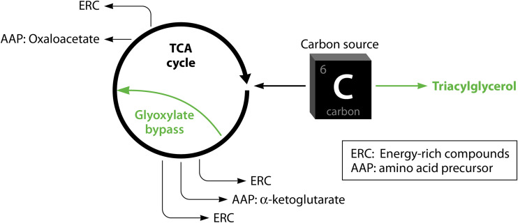
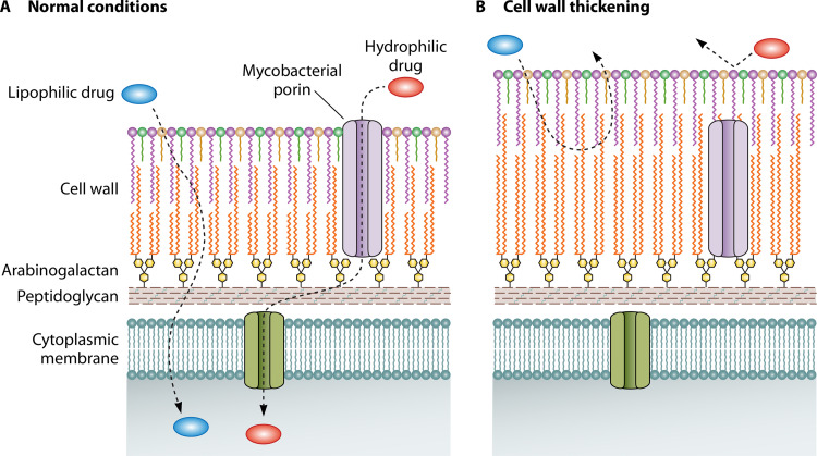
Successful treatment of tuberculosis (TB) can be hampered by Mycobacterium tuberculosis populations that are temporarily able to survive antibiotic pressure in the absence of drug resistance-conferring mutations, a phenomenon termed drug tolerance. We summarize findings on M. tuberculosis tolerance published in the past 20 years. Key M. tuberculosis responses to drug pressure are reduced growth rates, metabolic shifting, and the promotion of efflux pump activity. Metabolic shifts upon drug pressure mainly occur in M. tuberculosis's lipid metabolism and redox homeostasis, with reduced tricarboxylic acid cycle activity in favor of lipid anabolism. Increased lipid anabolism plays a role in cell wall thickening, which reduces sensitivity to most TB drugs. In addition to these general mechanisms, drug-specific mechanisms have been described. Upon isoniazid exposure, M. tuberculosis reprograms several pathways associated with mycolic acid biosynthesis. Upon rifampicin exposure, M. tuberculosis upregulates the expression of its drug target rpoB Upon bedaquiline exposure, ATP synthesis is stimulated, and the transcription factors Rv0324 and Rv0880 are activated. A better understanding of M. tuberculosis's responses to drug pressure will be important for the development of novel agents that prevent the development of drug tolerance following treatment initiation. Such agents could then contribute to novel TB treatment-shortening strategies.

Keywords: Mycobacterium tuberculosis; WhiB regulon; drug tolerance; efflux pumps; lipid metabolism; metabolic shifting; metabolic slowdown; mycobacterial cell wall; redox homeostasis; sigma factors.


Figures

FIG 1
FIG 1
Mechanisms for cross-tolerance in Mycobacterium tuberculosis. (A) Under nonstress conditions, cellular processes driving core metabolic pathways are unaffected. (B) Under drug pressure, homeostasis is affected by targeting core metabolic processes and toxic compounds, resulting in cellular damage and eventually cell death. (C) M. tuberculosis (Mtb) adapts to drug pressure through transcriptional and posttranscriptional regulatory mechanisms to develop a drug-tolerant state: metabolic slowdown reduces drug-induced cellular damage (1); metabolic processes are rerouted, and homeostasis is retained through the up- and downregulation of interweaved pathways (2); cell wall thickening limits drug entry into the cell and lowers drug activity (3); and the upregulation of efflux pumps lowers intracellular drug concentrations (4).
FIG 2
FIG 2
Metabolic shifting of carbon fluxes away from the TCA cycle. The promotion of FA synthesis through the upregulation of tgs1 is one strategy adopted by M. tuberculosis to limit carbon uptake in the TCA cycle by competing for mutual carbon sources. Reduced TCA cycle activity results in lower rates of turnover of alpha-ketoglutarate and oxaloacetate, both of which are metabolites required for amino acid synthesis. The upregulation of the glyoxylate bypass further limits the turnover of alpha-ketoglutarate.
FIG 3
FIG 3
Cell wall thickening. Cell wall thickening is a tolerance mechanism that results in the less efficient transport of lipophilic drugs across the mycobacterial cell wall. The transport of hydrophilic drugs is reduced as fewer porins now extend across the entire thickness of the cell wall.
FIG 4
FIG 4
Isoniazid (INH)-specific tolerance in Mycobacterium tuberculosis. (A) In the absence of tolerance, INH inhibits mycolic acid synthesis, resulting in insufficient mycolic acid production for growth. (B) Upon INH exposure, M. tuberculosis employs several tolerance mechanisms to overcome the INH-induced inhibition of mycolic acid synthesis: de novo-synthesized fatty acids and degraded high-molecular-weight TAGs fuel mycolic acid synthesis by acting as a source of mycolic acid precursors that enter mycolic acid synthesis (1), genes involved in mycolic acid synthesis (cmaA2, cmrA, mmaA2, mmaA3, mmaA4, umaA1, and fbpC) are upregulated (2), reduced carbon flow toward the TCA cycle leads to metabolic slowdown due to lowered ATP and protein synthesis (3), the promotion of mycolic acid synthesis and the reduced need for mycolic acids in light of reduced growth ensure sufficient mycolic acid production (4), and M. tuberculosis decreases the expression of NADH dehydrogenase to avoid the further accumulation of NAD+ when NAD+ is accumulated upon increased production of fatty acids and mycolic acids and reduced TCA cycle activity (5).
FIG 5
FIG 5
Bedaquiline (BDQ)-specific tolerance in Mycobacterium tuberculosis. The effects of BDQ exposure in the absence of tolerance (red) and the presence of tolerance (green) consist of multiple adaptations: efflux pump activity that occurs even in the nontolerant state and may be increased in the tolerant state (A), metabolic slowdown to reduce ATP dependency (B), and promoting ATP synthesis to overcome the ATP shortage induced by BDQ, either directly through the overexpression of genes encoding subunits of the ATP synthase machinery or indirectly by the stimulation of oxidative phosphorylation (C). The latter can be performed by increasing the synthesis of components used during oxidative phosphorylation (e.g., cytochromes) and/or by promoting trehalose metabolism and TCA cycle activity to increase the production of the energy-rich metabolites that power oxidative phosphorylation and generate the proton motive force that is required for ATP synthesis.

References

    1. WHO. 2019. Global tuberculosis report 2019. WHO, Geneva, Switzerland: https://www.who.int/tb/publications/global_report/en/.
    1. WHO. 2018. Global tuberculosis report 2018. WHO, Geneva, Switzerland: https://www.who.int/tb/publications/global_report/en/.
    1. Grace AG, Mittal A, Jain S, Tripathy JP, Satyanarayana S, Tharyan P, Kirubakaran R. 2018. Shortened treatment regimens versus the standard regimen for drug-sensitive pulmonary tuberculosis. Cochrane Database Syst Rev 12:CD012918. doi:10.1002/14651858.CD012918. - DOI - PMC - PubMed
    1. Yihunie Akalu T, Muchie KF, Alemu Gelaye K. 2018. Time to sputum culture conversion and its determinants among multi-drug resistant tuberculosis patients at public hospitals of the Amhara Regional State: a multicenter retrospective follow up study. PLoS One 13:e0199320. doi:10.1371/journal.pone.0199320. - DOI - PMC - PubMed
    1. Gillespie SH, Crook AM, McHugh TD, Mendel CM, Meredith SK, Murray SR, Pappas F, Phillips PP, Nunn AJ, REMoxTB Consortium. 2014. Four-month moxifloxacin-based regimens for drug-sensitive tuberculosis. N Engl J Med 371:1577–1587. doi:10.1056/NEJMoa1407426. - DOI - PMC - PubMed

Publication types

MeSH terms