. 2021 Jan 4;131(1):e130740.
doi: 10.1172/JCI130740.

Galectin-7 downregulation in lesional keratinocytes contributes to enhanced IL-17A signaling and skin pathology in psoriasis

Affiliations

Galectin-7 downregulation in lesional keratinocytes contributes to enhanced IL-17A signaling and skin pathology in psoriasis

Hung-Lin Chen et al. J Clin Invest. .

Abstract

Psoriasis is a chronic inflammatory skin disease characterized by inflammatory cell infiltration, as well as hyperproliferation of keratinocytes in skin lesions, and is considered a metabolic syndrome. We found that the expression of galectin-7 is reduced in skin lesions of patients with psoriasis. IL-17A and TNF-α, 2 cytokines intimately involved in the development of psoriatic lesions, suppressed galectin-7 expression in human primary keratinocytes (HEKn cells) and the immortalized human keratinocyte cell line HaCaT. A galectin-7 knockdown in these cells elevated the production of IL-6 and IL-8 and enhanced ERK signaling when the cells were stimulated with IL-17A. Galectin-7 attenuated IL-17A-induced production of inflammatory mediators by keratinocytes via the microRNA-146a/ERK pathway. Moreover, galectin-7-deficient mice showed enhanced epidermal hyperplasia and skin inflammation in response to intradermal IL-23 injection. We identified fluvastatin as an inducer of galectin-7 expression by connectivity map analysis, confirmed this effect in keratinocytes, and demonstrated that fluvastatin attenuated IL-6 and IL-8 production induced by IL-17A. Thus, we validate a role of galectin-7 in the pathogenesis of psoriasis, in both epidermal hyperplasia and keratinocyte-mediated inflammatory responses, and formulate a rationale for the use of statins in the treatment of psoriasis.

Keywords: Autoimmune diseases; Autoimmunity; Dermatology; Skin.


Conflict of interest statement

Conflict of interest: FTL, HLC, and PCC are inventors on patent application numbers US 16/653,986 and Taiwan 108137064.

Figures

Figure 1
Figure 1. The levels of galectin-7 are low in the epidermal keratinocytes of patients with psoriasis.
(A) Representative immunohistochemical (IHC) staining pictures of galectin-7 in skin sections from a healthy control and a psoriatic lesion. Scale bars: 100 μm. (B) Quantification of IHC staining of galectin-7 in sections of normal skin from healthy controls (n = 75) and sections of lesional skin from patients with psoriasis (n = 27). Quantification of staining was performed by computer-assisted methods as described in Methods. The y axis indicates arbitrary numbers representing mean intensity across an area of the epidermis (InteDen/area). (C) IHC staining of galectin-7 in lesional skin and nonlesional skin pair. (D) Quantification of galectin-7 staining of paired lesional and nonlesional skin from the same patients (n = 10 pairs). (E) IHC staining of galectin-7 in sections from intradermally IL-23–injected and PBS-injected mouse skin. Scale bars: 100 μm. (F) Quantification of galectin-7 staining of PBS-injected (n = 6) and IL-23–injected mouse skin (n = 13). (G) Immunoblot analysis of galectin-7 levels in HaCaT and HEKn cells stimulated with the indicated cytokines (IFN-γ, LPS, TNF-α, IL-23, or IL-17A). The concentrations of IFN-γ and LPS used were 100 ng/mL and 50 μg/mL, respectively. The concentrations of TNF-α, IL-23, and IL-17A used were as indicated in the figure. Cells were treated with the cytokines for 2 days, and cell lysates were prepared for immunoblot analysis. GAPDH served as a loading control. The intensity of the galectin-7 band was quantified by densitometry and normalized to GAPDH and control samples. The number below each band represents the relative galectin-7 amount. Statistical analyses were performed by unpaired (B and F) and paired (D) Student’s t test. *P < 0.05, ***P < 0.001.
Figure 2
Figure 2. Downregulation of galectin-7 in keratinocytes causes an elevated production of the proinflammatory cytokines IL-6 and IL-8 in response to IL-17A stimulation.
(A and B) Galectin-7–knockdown HaCaT cell lines (sh-1, sh-2, sh-3, and sh-4) and controls were incubated with or without IL-17A for 2 days, and IL-6 and IL-8 concentrations in the supernatants were measured by ELISA. All the experiments included 3 biological replicates. (C and D) HEKn cells were transfected with siRNA to knock down galectin-7 and then incubated with or without IL-17A for 2 days. The IL-6 and IL-8 concentrations in the supernatants were measured by ELISA. Three independent biological replicates were performed for the ELISA analysis. The results are presented as mean ± SD. For statistical analysis, 2-way ANOVA with Tukey’s multiple-comparison test was performed. Each shRNA- or siRNA-treated cell line was compared with its corresponding control (V, vector; si-NC, negative control siRNA) for both untreated and IL-17–treated groups. The shRNAs and siRNAs for the knockdown of galectin-7 expression were as described in Methods. *P < 0.05, **P < 0.01.
Figure 3
Figure 3. MiR-146a is upregulated in the epidermis of psoriatic lesions; this miRNA promotes production of the proinflammatory cytokines IL-6 and IL-8.
(A) MiR-146a in the epidermis of normal and psoriatic skin was detected by RNA in situ hybridization assay. Scale bars: 100 μm. (B) The miR-146a level in HaCaT cells treated with IL-17A was quantified by real-time PCR. (C and D) A miR-146a–overexpressing vector (pmiR-146a) and a scrambled control hairpin in pCDH-CMV-MCS-EF1-copGFP (pmiR) from SBI (System Biosciences) were used to generate stable miR-146a–overexpressing and control cell clones, respectively. The secretion of cytokines (IL-6 and IL-8) by HaCaT cells stably transfected with pmiR or pmiR-146a vectors was measured 2 days after stimulation with 25 or 100 ng/mL IL-17A. Three independent biological replicates were performed for the real-time PCR analysis. All results are presented as mean ± SD. For statistical analysis, unpaired Student’s t test (B) or 2-way ANOVA with Tukey’s multiple-comparison test (C and D) was performed. **P < 0.01, ***P < 0.001.
Figure 4
Figure 4. Reduced galectin-7 expression and miR-146a overexpression promote ERK1 and ERK2 signaling pathways triggered by IL-17A.
(A) Galectin-7–knockdown HaCaT cells and control cells were treated with IL-17A for 5 minutes, and cell lysates were analyzed by immunoblotting. Total ERK1, ERK2, NF-κB, and IκBα and their phosphorylated forms were detected with the corresponding antibodies. (B) HaCaT cells stably transfected with pmiR (control vector) or pmiR-146a were treated with IL-17A for 5 minutes. Immunoblotting was performed as described in A. Protein quantification data on phospho-ERK1 (p-ERK1) and phospho-ERK2 (p-ERK2) were normalized to the control group in A and the miR group in B. Data on total protein levels, levels of phosphorylated NF-κB and IκBα, and total protein levels of ERK1 and ERK2 were normalized to the control (0 minutes).
Figure 5
Figure 5. Galectin-7–deficient mice exhibit hyperproliferative keratinocytes and increased leukocyte infiltration at the intradermally IL-23–injected sites, as compared with their littermate controls.
(A) H&E staining of ear sections from WT or galectin-7–deficient (knockout; KO) mice injected intradermally with PBS or IL-23 every other day for 15 days. Scale bars: 50 μm. (B) Ear thickness of WT and galectin-7–deficient (KO) mice was measured before each intradermal injection, and measurements were taken at the center of the ears (WT/PBS, n = 5; WT/IL-23, n = 18; KO/PBS, n = 5; KO/IL-23, n = 19). For statistical analysis, ear thickness of KO/IL-23 was compared with that in the corresponding WT/IL-23 group at each time point. (C) Epidermal thicknesses of WT and KO mice from H&E-stained sections as described in A obtained on day 15 from the same mice as described in B. For each tissue section, 3 measurements were taken. (D) Leukocytes were counted in ×400 magnified visual fields of tissue sections from IL-23–injected mice using the cell counting module in ImageJ software (WT, n = 7; KO, n = 5). All results are presented as mean ± SD. For statistical analysis, 2-way ANOVA with Tukey’s multiple-comparison test (B and C) or unpaired Student’s t test (D) was performed. **P < 0.01, ***P < 0.001.
Figure 6
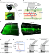
Figure 6. Two-photon deep-tissue visualization of EGFP–galectin-7 expression in transgenic mice.
(A) Detection of green fluorescent signal on the whole-body surface. (B) Schematic representation of the 2-photon image analysis of deep tissue in the ear. (C) Three-dimensional images of the view of x-y axis and z axis planes. (D) Immunoblot analysis of galectin-7–GFP expression from tissues of EGFP–galectin-7 transgenic mouse. (E) Flow cytometry analysis of bone marrow cells purified from WT and EGFP–galectin-7 transgenic mice. (F) Affinity purification of EGFP–galectin-7 fusion protein from the lysates of skin samples from EGFP–galectin-7 transgenic mice, followed by immunoblot analysis.
Figure 7
Figure 7. Fluvastatin increases galectin-7 levels in keratinocytes, suppresses production of proinflammatory cytokines, and attenuates IL-23–induced epidermal thickness.
(A and B) Real-time PCR analysis of the mRNA expression of galectin-7 and S100A7 in HaCaT cells treated with methylprednisolone (10.6 μM), tobramycin (8.6 μM), fluvastatin (9.2 μM), pempidine (13 μM), or vehicle control (DMSO) for 24 hours. The relative fold changes were calculated using the ΔΔCt method; data from all the samples were normalized to the control sample, and GAPDH served as an endogenous control. (C and D) Production of IL-6 and IL-8 by HaCaT cells treated for 2 days with fluvastatin with or without IL-17A (200 ng/mL) was measured by ELISA. For statistical analysis, each group was compared with control group (first bar: group without IL-17A, DMSO, and fluvastatin). (E) Immunoblotting analysis of galectin-7 in keratinocytes treated with TNF-α or IL-17A with or without fluvastatin or DMSO. (F) Ear thicknesses of different groups — PBS (n = 3), IL-23 (n = 3), PBS + vehicle (n = 5), IL-23 + vehicle (n = 5), PBS + fluvastatin (n = 3), IL-23 + fluvastatin (n = 5), PBS + pravastatin (n = 4), IL-23 + pravastatin (n = 5) — of mice subjected to intradermal injections of IL-23 or PBS, as described in Figure 5, and treated with fluvastatin (blue line), pravastatin (green line), or saline. For statistical analysis, either 1-way (A and B) or 2-way (C, D, and F) ANOVA with Tukey’s multiple-comparison test was used. Three independent biological replicates were performed for real-time PCR and ELISA analyses. The results (adjusted P values) of group pairs are annotated on the graph (IL-23 + fluvastatin group vs. corresponding IL-23 + vehicle group indicated in gray; IL-23 + fluvastatin group vs. corresponding IL-23 + pravastatin group indicated in green). *P < 0.05, **P < 0.01, ***P < 0.001.

Similar articles

Cited by

References

    1. Polyak K, Xia Y, Zweier JL, Kinzler KW, Vogelstein B. A model for p53-induced apoptosis. Nature. 1997;389(6648):300–305. doi: 10.1038/38525. - DOI - PubMed
    1. Bernerd F, Sarasin A, Magnaldo T. Galectin-7 overexpression is associated with the apoptotic process in UVB-induced sunburn keratinocytes. Proc Natl Acad Sci U S A. 1999;96(20):11329–11334. doi: 10.1073/pnas.96.20.11329. - DOI - PMC - PubMed
    1. Kuwabara I, et al. Galectin-7 (PIG1) exhibits pro-apoptotic function through JNK activation and mitochondrial cytochrome c release. J Biol Chem. 2002;277(5):3487–3497. doi: 10.1074/jbc.M109360200. - DOI - PubMed
    1. Magnaldo T, Bernerd F, Darmon M. Galectin-7, a human 14-kDa S-lectin, specifically expressed in keratinocytes and sensitive to retinoic acid. Dev Biol. 1995;168(2):259–271. doi: 10.1006/dbio.1995.1078. - DOI - PubMed
    1. Gendronneau G, et al. Galectin-7 in the control of epidermal homeostasis after injury. Mol Biol Cell. 2008;19(12):5541–5549. doi: 10.1091/mbc.e08-02-0166. - DOI - PMC - PubMed

Publication types