. 2020 Oct 15;5(20):e141523.
doi: 10.1172/jci.insight.141523.

A porcine model of phenylketonuria generated by CRISPR/Cas9 genome editing

Affiliations

A porcine model of phenylketonuria generated by CRISPR/Cas9 genome editing

Erik A Koppes et al. JCI Insight. .

Abstract

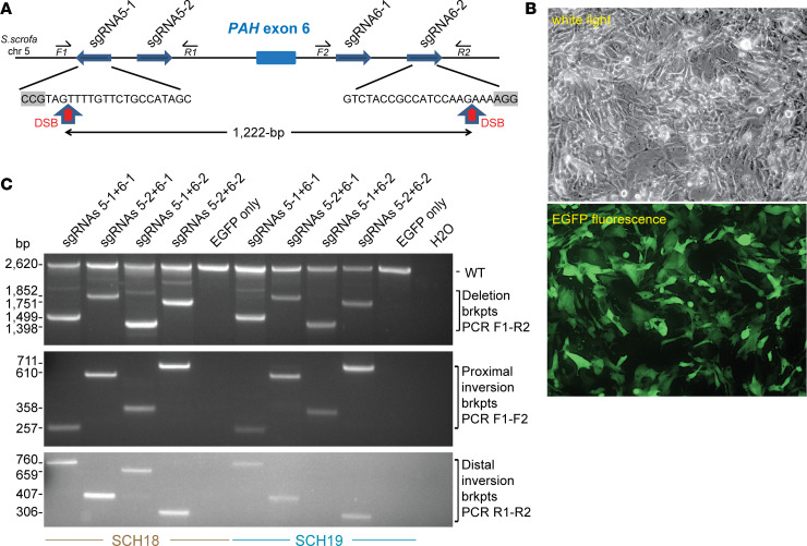
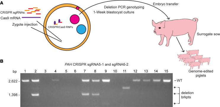
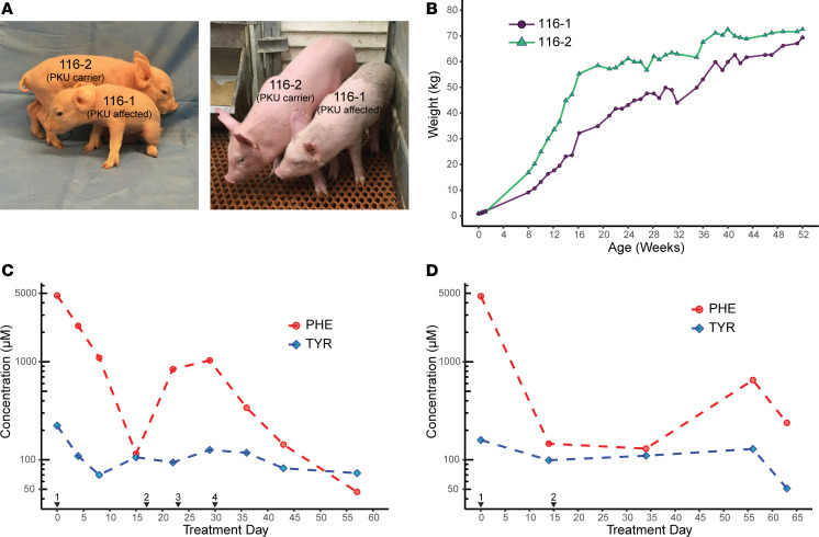
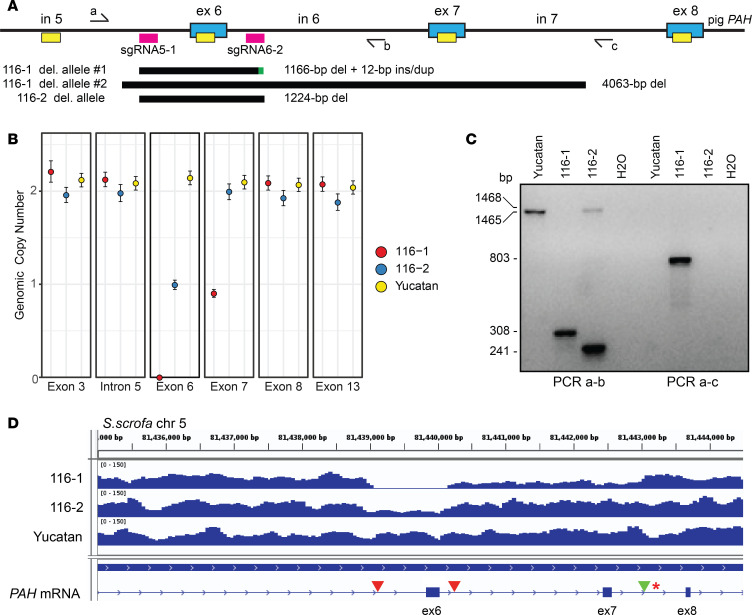
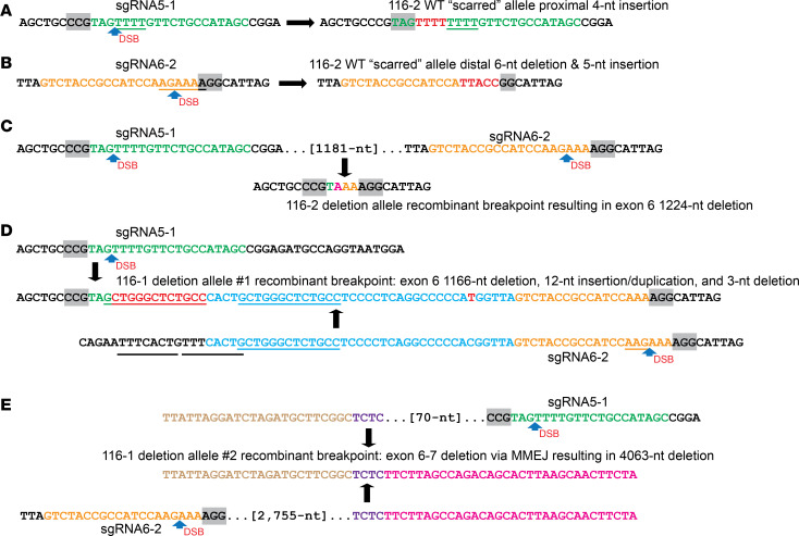
Phenylalanine hydroxylase-deficient (PAH-deficient) phenylketonuria (PKU) results in systemic hyperphenylalaninemia, leading to neurotoxicity with severe developmental disabilities. Dietary phenylalanine (Phe) restriction prevents the most deleterious effects of hyperphenylalaninemia, but adherence to diet is poor in adult and adolescent patients, resulting in characteristic neurobehavioral phenotypes. Thus, an urgent need exists for new treatments. Additionally, rodent models of PKU do not adequately reflect neurocognitive phenotypes, and thus there is a need for improved animal models. To this end, we have developed PAH-null pigs. After selection of optimal CRISPR/Cas9 genome-editing reagents by using an in vitro cell model, zygote injection of 2 sgRNAs and Cas9 mRNA demonstrated deletions in preimplantation embryos, with embryo transfer to a surrogate leading to 2 founder animals. One pig was heterozygous for a PAH exon 6 deletion allele, while the other was compound heterozygous for deletions of exon 6 and of exons 6-7. The affected pig exhibited hyperphenylalaninemia (2000-5000 μM) that was treatable by dietary Phe restriction, consistent with classical PKU, along with juvenile growth retardation, hypopigmentation, ventriculomegaly, and decreased brain gray matter volume. In conclusion, we have established a large-animal preclinical model of PKU to investigate pathophysiology and to assess new therapeutic interventions.

Keywords: Amino acid metabolism; Genetic diseases; Genetics; Metabolism; Mouse models.


Conflict of interest statement

Conflict of interest: The authors have declared that no conflict of interest exists.

Figures

Figure 1
Figure 1. Strategy and optimization in vitro for CRISPR/Cas9 gene editing of the pig PAH gene.
(A) Strategy showing paired CRISPR sgRNA targeting to generate deletion or inversion of PAH exon 6. Key: thick directional blue arrows, sgRNAs targeting the sense or complementary strand of intron 5 or intron 6; small directional arrows, forward (F) or reverse (R) deletion and inversion genotyping PCR primers; red vertical arrows, DSB sites in optimal sgRNA5-1 and sgRNA6-2 at a position 3 nt 5′ of the PAM motif (gray boxes). (B) Representative images (white light and EGFP epifluorescence, original magnification, ×10) for transfection of the SCH18 line with plasmids encoding Cas9, CRISPR sgRNA5-1 + sgRNA6-2, and EGFP. (C) Deletion and inversion PCR assays to detect exon 6 genome editing in SCH lines. Top: Deletion PCR assay with genotyping primers F1 and R2, for SCH18 and SCH19 cells transfected with pairs of sgRNA vectors plus EGFP or EGFP only as a control. Deletion breakpoints (brkpts) are only amplified in cells transfected with Cas9/sgRNA vectors whereas the WT band is detected in all samples. Middle and bottom: Inversion PCR assays for proximal and distal inversion breakpoints using primer pairs F1 and F2 or R1 and R2, respectively. Inversion breakpoints are detected in cells transfected with pairs of Cas9/sgRNA vectors indicating robust genome editing and NHEJ repair using the inverted exon 6 segment to bridge the DSB sites.
Figure 2
Figure 2. Zygote RNA injection, blastocyst screening, and embryo transfer to derive genome-edited pigs.
(A) Strategy for in vivo genome editing of pig zygotes with injection of CRISPR sgRNAs and Cas9 mRNA into fertilized zygotes, and growth for 5–7 days in vitro, followed by DNA analysis of blastocysts or embryo transfer to a surrogate. RNP,. ribonucleoprotein. (B) Deletion PCR confirmation of gene editing of PAH exon 6 in a representative set of 15 pig blastocysts. PCR genotyping (as for Figure 1C) detects the WT band in 10 of these individual blastocysts while the expected deletion breakpoint (brkpt) band (1398 bp) is also detected in blastocysts 2, 7, and 8. Variant bands representing different deletion sizes are detected in blastocysts 9 and 11. Of a total of 57 blastocysts analyzed from 2 embryo transfers, 40 produced a PCR band(s), and of these 19 showed deletion alleles.
Figure 3
Figure 3. Phenotypes of affected and heterozygous PAH-deficient PKU pigs.
(A) The PKU animal 116-1 had juvenile growth retardation and hypopigmentation compared with her heterozygous littermate 116-2. (B) Growth chart of affected pig 116-1 and heterozygous pig 116-2 pigs, demonstrating growth retardation in the former followed by catch-up growth toward 1 year of age. (C) Dietary treatment (trial 1) of the affected PKU pig results in reduction of blood Phe levels into therapeutic range and normalization of the Phe/Tyr ratio. Black arrowheads indicate days on which diet was changed to (1) initiation of Phe washout with 100% Phe-free chow, followed by adjustment of diet to (2) 50-50 normal to Phe-free chow; (3) 75-25 Phe-free to normal chow; and (4) 85-15 Phe-free to normal chow. Red circles, Phe levels; blue diamonds, Tyr levels. Amino acid concentration (μM, shown on a log10 scale) was assayed from blood plasma before and during dietary treatment as determined by HPLC. (D) Two-step dietary treatment (trial 2) of the affected PKU pig results in normalization of blood Phe levels and of the Phe/Tyr ratio. Details are as for C with (1) initiation of Phe washout with 100% Phe-free chow, followed by (2) adjustment of diet to 85-15 Phe-free to normal chow.
Figure 4
Figure 4. Molecular characterization of PAH deletion alleles in the pig PKU founder animals.
(A) Schematic of CRISPR/Cas9 targeting of the PAH locus and detection of genome-edited alleles by ddPCR and long-range genomic PCR. Key: blue boxes, PAH exons (ex) with intervening introns (in); pink boxes, CRISPR sgRNA sites; yellow boxes, ddPCR amplicons; half arrows, genotyping primers a-c; black bars, regions deleted; green bar, insertion/duplication. (B) Genomic copy number across the PAH locus as determined by normalization of copies/μL to the single copy gene GAPDH (all amplitude plots are shown in Supplemental Figure 6), indicating heterozygosity at PAH exon 6 in 116-2 and homozygous exon 6 deletion along with heterozygous deletion of exon 7 in 116-1. Error bars were calculated based on the Poisson distribution. (C) PCR genotyping using primer pairs a-b to amplify a WT (1465 bp) fragment in Yucatan, a scarred allele (1468 bp) in 116-2, and exon 6 deletions in 116-1 and 116-2 (308 and 241 bp, respectively) and primer pair a-c to amplify the larger exon 6–7 deletion allele from 116-1 (803 bp). (D) Whole-genome sequencing (WGS) read depth coverage across the PAH exon 6–8 region in the founding PKU pigs and a Yucatan control. Note the absence of reads within exon 6 and reduced reads extending to intron 7 in the compound heterozygous deletion animal 116-1 and the reduced number of reads in exon 6 for the heterozygous animal 116-2. Red arrowheads mark the CRISPR sgRNA target sites, and the green arrowhead indicates the exon 6–7 distal breakpoint present on the larger 116-1 PAH deletion allele. The red asterisk represents an SNP linked to breakpoint reads used to infer the parental origin of the larger 116-1 deletion allele.
Figure 5
Figure 5. DNA repair mechanisms for deletion and scarred gene-edited PAH alleles.
(A and B) The intact (WT-size) allele for 116-2 (heterozygote) demonstrates gene editing (“scarring”) at each sgRNA target site. Inaccurate NHEJ mechanisms lead to (A) the insertion of 4 T nucleotides (red) on DNA repair of a DSB at the sgRNA5-1 position, while (B) DNA repair of a DSB at the sgRNA6-2 position leads to both deletion of 6 nt (AGAAAA) including the first nucleotide of the PAM sequence and insertion of 5 nt of novel sequence (red; TTACC), respectively. (C) The deletion allele for 116-2 (heterozygote) arose by DNA repair of DSBs at each sgRNA position with loss of the intervening PAH exon 6 segment. Two extra nucleotides were deleted as part of the mechanism either due to noncanonical positioning of each DSB by CRISPR/Cas9, or, perhaps most likely, the inaccurate NHEJ mechanism of DNA repair. (D) A complex NHEJ mechanism accounts for the exon 6 recombinant deletion allele 1 in 116-1 (affected PKU pig). CRISPR/Cas9-induced DSBs at the canonical positions 3 nt 5′ of the PAM sequences for the sgRNA5-1 and sgRNA6-2 sgRNAs lead to a series of potential events that can be described by 2 alternative DNA repair mechanisms (see main text). Descriptively, the recombinant chromosome is characterized by a 1166 bp deletion starting at the sgRNA5-1 canonical position and with insertion at the deletion breakpoint of a novel 12 nt sequence apparently derived from an adjacent sequence to generate a 13 nt duplication (this and an adjacent 8 nt tandem repeat in the distal parental sequence are underlined). The distal deletion breakpoint is localized between the 13 nt duplication at a position 56 nt upstream of the canonical position for a DSB at sgRNA6-2, with a 3 nt deletion (AAG) at the latter DSB. A single SNP (C→T) arising in the mutant allele is also detected. (E) A complex MMEJ mechanism accounts for the larger exon 6–7 deletion allele 2 in 116-1 (PKU). DNA repair of the paired DSBs at sgRNA5-1 and sgRNA6-2 led to a recombinant breakpoint at a 4 nt (TCTC, purple) sequence of patchy homology located 70 nt 5′ and 2755 nt 3′ of the sgRNA target sites, respectively, resulting in a 4063 nt deletion.

References

    1. Donlon J, Sarkissian C, Levy H, Scriver CR. Hyperphenylalaninemia: phenylalanine hydroxylase deficiency. In: Valle D, Antonarakis S, Ballabio A, Beaudet A, Mitchell GA, eds. The Online Metabolic and Molecular Bases of Inherited Disease. McGraw-Hill; Accessed August 10, 2020. https://ommbid-mhmedical-com.pitt.idm.oclc.org/content.aspx?bookid=2709&....
    1. Scriver C, Kaufman S. Hyperphenylalaninemia: phenylalanine hydroxylase deficiency. In: Beaudet A, et al. eds. The Metabolic & Molecular Bases of Inherited Disease. McGraw-Hill; 2001:1667–1724.
    1. Vockley J, et al. Phenylalanine hydroxylase deficiency: diagnosis and management guideline. Genet Med. 2014;16(2):188–200. doi: 10.1038/gim.2013.157. - DOI - PubMed
    1. Scriver CR, et al. PAHdb: a locus-specific knowledgebase. Hum Mutat. 2000;15(1):99–104. doi: 10.1002/(SICI)1098-1004(200001)15:1<99::AID-HUMU18>3.0.CO;2-P. - DOI - PubMed
    1. Williams RA, Mamotte CD, Burnett JR. Phenylketonuria: an inborn error of phenylalanine metabolism. Clin Biochem Rev. 2008;29(1):31–41. - PMC - PubMed

Publication types