. 2020 Oct 14;10(1):17225.
doi: 10.1038/s41598-020-73945-w.

Lacrimal gland excision in male and female mice causes ocular pain and anxiety-like behaviors

Affiliations

Lacrimal gland excision in male and female mice causes ocular pain and anxiety-like behaviors

Neal E Mecum et al. Sci Rep. .

Abstract

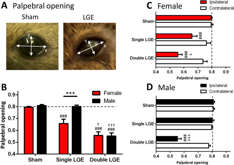
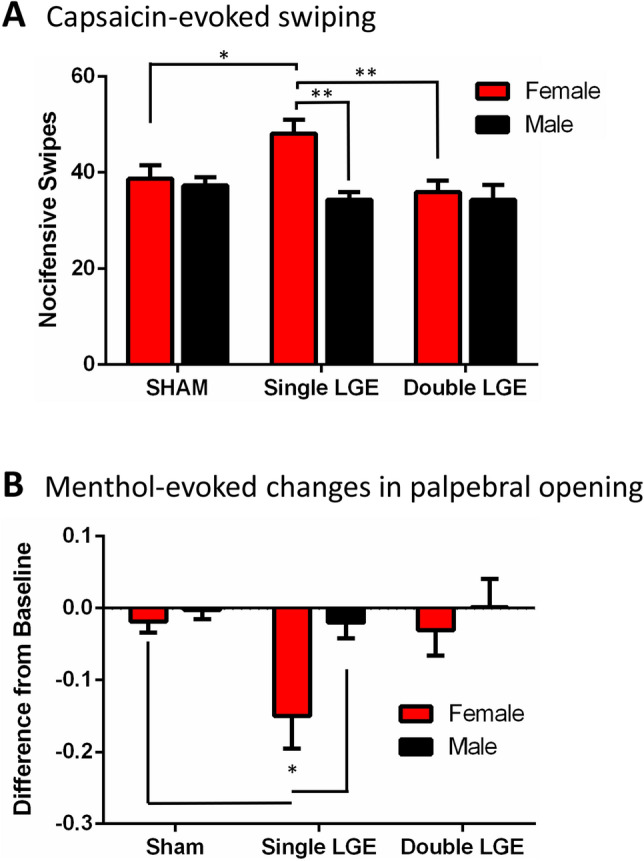
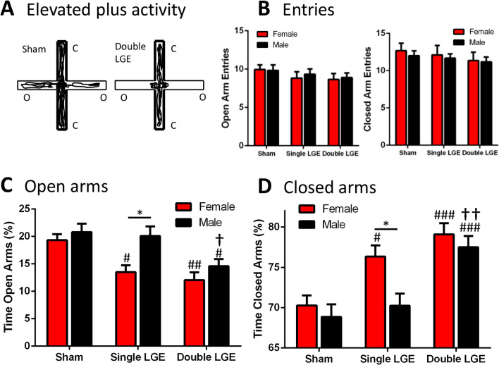
Lacrimal gland excision (LGE) induced dry eye produces more severe corneal damage in female mice, yet signs of LGE-induced ocular pain and anxiety in male and female mice have not been characterized. Excision of either the extraorbital gland (single LGE), or both the extraorbital and intraorbital glands (double LGE) was performed in male and female C57BL/6J mice to induce moderate and severe dry eye. Ongoing pain was assessed by quantifying palpebral opening and evoked nociceptive responses after corneal application of capsaicin and menthol. The open-field and plus maze were used to assess anxiety. Single LGE caused a reduction in palpebral opening and an increase in capsaicin and menthol-evoked responses only in female mice. Furthermore, single LGE produced signs of increased anxiety in female but not male mice. Overall, female mice appear more susceptible to signs of ocular pain, irritation, and anxiety in response to aqueous tear deficiency.


Conflict of interest statement

The authors declare no competing interests.

Figures

Figure 1
Figure 1
Palpebral opening following lacrimal gland excision in female and male mice. (A) Representative photos showing an eye from a sham animal (left) and from an animal following double LGE (right). The palpebral opening was calculated as the ratio Y/X. (B) Single LGE caused a significant reduction in palpebral opening only in female mice, whereas double LGE produced comparable reductions in the palpebral opening between female and male mice. (C) In female mice, both single and double LGE resulted in a significant reduction in palpebral opening in the eye ipsilateral to gland excision. (D)In male mice, only double LGE caused a significant reduction in palpebral opening on the side ipsilateral to gland excision. Dashed line represents naïve palpebral opening score. n = 12/treatment group. *p < 0.05, **p < 0.01, ***p < 0.001. ###p < 0.001 compared to sham of the same sex; p < 0.05, †††p < 0.001 compared to single LGE of the same sex.
Figure 2
Figure 2
The effect of topical anesthetics on palpebral opening in double lacrimal gland excision treated animals. Corneal application of (A) tetracaine, and (B) proparacaine increased the palpebral opening for 5 min before gradually returning to baseline values. (C) Corneal application of oxybuprocaine produced a more prolonged anesthesia, with the palpebral opening returning to baseline after 30 min. (D) Corneal application of artificial tears had no effect on eye closure 5 min post application. BSL, baseline. n = 6 for each drug treatment group (3 females, 3 males). Dashed line represents naïve palpebral opening score. *p < 0.05, **p < 0.01, ***p < 0.001; #p < 0. 0001 compared to baseline; p < 0.0001 compared to artificial tears.
Figure 3
Figure 3
Ocular sensitivity to corneal application of capsaicin and menthol. (A) The number of nocifensive eye wipes after application of capsaicin was significantly greater only in female animals that had undergone single LGE. N = 11–12/treatment group. (B) After a baseline measurement, the palpebral opening was quantified 5-min post application of menthol. Menthol caused a decrease in the palpebral opening only in female mice with single LGE. n = 12/treatment group. *p < 0.05, **p < 0.01.
Figure 4
Figure 4
Locomotor activity following lacrimal gland excision. (A, B) Total distance traveled in female and male mice. Double LGE decreased the distance traveled in both female and male mice compared to sham treated controls. (C, D) Total number of vertical rears in female and male mice. In female mice, single and double LGE caused a significant reduction in total rears compared to sham controls. In male mice, only double LGE decreased the number of rears. (E, F) Total vertical rearing time for female and male mice. In female mice, single and double LGE caused a significant drop in vertical rearing time compared to sham controls. In male mice, only double LGE caused a significant drop in vertical rearing time compared to sham controls. n = 16–18/treatment group. *p < 0.05, **p < 0.01.
Figure 5
Figure 5
The effect of lacrimal gland excision on elevated plus maze behavior. (A) Representative examples of elevated plus maze activity in a sham mouse (left) and in a double LGE treated mouse (right). O, open arms; C, closed arms. (B) The number of open (left) and closed (right) arm entries were similar between treatment groups. (C) Percentage of time female and male mice spent in the open arms. Single and double LGE decreased the time female mice spent in the open arms compared to female sham animals. In male mice, only double LGE caused a significant decrease in open arm time. (D) Percentage of time female and male mice spent in the closed arms. Single LGE increased time in the closed arms only in female mice, whereas double LGE increased time spent in the closed arms in both female and male mice. n = 17–18/treatment group. *p < 0.05, **p < 0.01, ***p < 0.001; #p < 0.05, ##p < 0.01, ###p < 0.001 compared to sham of the same sex; p < 0.05, ††p < 0.01 compared to single LGE of the same sex.
Figure 6
Figure 6
Corneal innervation two weeks after sham and single LGE in male and female Nav1.8-cre;tdTomato mice. (A) Representative image of the cornea showing nerve innervation for different regions of interest as well as layers including subbasal and intraepithelial terminals. Scale bar 20 μm. (B) Representative images showing the concentric rings placed onto the corneal regions of interest for use in the Sholl analysis for the subbasal nerve plexus (top left) and intraepithelial terminals (top right). Scale bar 20 μm. The number of Sholl intersections for the subbasal nerve plexus (bottom left) and intraepithelial terminals (bottom right) in female and male mice after sham and single LGE. (C) Pixel analysis of the subbasal nerve plexus (left) and intraepithelial terminals (right). (D) Tear measurements conducted 1-week after sham and single LGE. Single LGE reduced tear volume when compared to sham surgery in both female and male animals and no sex differences were observed. n = 6–8/treatment group. ***p < 0.001 versus sham operated controls.

References

    1. Meng ID, Kurose M. The role of corneal afferent neurons in regulating tears under normal and dry eye conditions. Exp. Eye Res. 2013;117:79–87. doi: 10.1016/j.exer.2013.08.011. - DOI - PMC - PubMed
    1. Dartt DA. Neural regulation of lacrimal gland secretory processes: relevance in dry eye diseases. Prog. Retin. Eye Res. 2009;28:155–177. doi: 10.1016/j.preteyeres.2009.04.003. - DOI - PMC - PubMed
    1. Tiffany J. The normal tear film. Dev. Ophthalmol. 2008;41:1–20. doi: 10.1159/000131066. - DOI - PubMed
    1. Li M, Gong L, Sun X, Chapin WJ. Anxiety and depression in patients with dry eye syndrome. Curr. Eye Res. 2011;36:1–7. doi: 10.3109/02713683.2010.519850. - DOI - PubMed
    1. Tian YJ, et al. Epidemiologic study of dry eye in populations equal or over 20 years old in Jiangning District of Shanghai. Chinese J. Ophthalmol. 2009;45:486–491. - PubMed

Publication types