. 2020 Oct 14;10(1):17257.
doi: 10.1038/s41598-020-74176-9.

Long-acting antibody ligand mimetics for HER4-selective agonism

Affiliations

Long-acting antibody ligand mimetics for HER4-selective agonism

Lu Shan et al. Sci Rep. .

Abstract

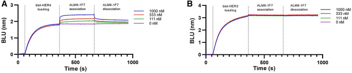
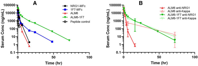
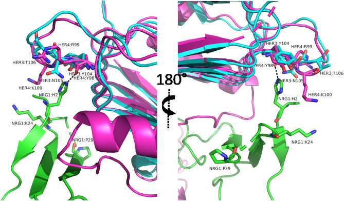
Neuregulin protein 1 (NRG1) is a large (> 60-amino-acid) natural peptide ligand for the ErbB protein family members HER3 and HER4. We developed an agonistic antibody modality, termed antibody ligand mimetics (ALM), by incorporating complex ligand agonists such as NRG1 into an antibody scaffold. We optimized the linker and ligand length to achieve native ligand activity in HEK293 cells and cardiomyocytes derived from induced pluripotent stem cells (iPSCs) and used a monomeric Fc-ligand fusion platform to steer the ligand specificity toward HER4-dominant agonism. With the help of selectivity engineering, these enhanced ALM molecules can provide an antibody scaffold with increased receptor specificity and the potential to greatly improve the pharmacokinetics, stability, and downstream developability profiles from the natural ligand approach. This ligand mimetic design and optimization approach can be expanded to apply to other cardiovascular disease targets and emerging therapeutic areas, providing differentiated drug molecules with increased specificity and extended half-life.


Conflict of interest statement

All authors are or were employees of AstraZeneca at the time the research was performed, with stock ownership and/or options in the company.

Figures

Figure 1
Figure 1
ALM design and characterization. (A) Unliganded structure of HER4 receptor ECD (PDB ID: 2AHX) shows the separation between domain 1 and domain 2 and a buried dimerization arm (blue). (B) Structure of neuregulin-bound HER4 (PBD ID: 3U7U) shows that NRG1 engage a receptor conformational change to form a binding interface with domains 1 and 2. Vertical 90-degree rotation shows the extended dimerization arm (blue). (C) Structural model of ALM designs, using the b12 antibody as the scaffold (PBD ID: 1HZH) and NRG1 (PDB ID: 3U7U). (D) The variable heavy-chain CDR3 loop was replaced with ligand sequences. (E) SEC-MALS analysis confirmed homogeneity of ALM6 after protein A chromatography purification, with the molecular size calculated from MALS. (F) SEC traces of ALM6 at 0.5 mg/mL (solid) and at 10 mg/mL (dashed). (G) HER2/HER4 dual-expressing HEK293 cell-based luciferase reporter assay showed a gradual increase in receptor activation from ALM3, ALM4, ALM5, and ALM6. (H) Phospho-AKT activation was measured on iPSC-derived cardiomyocytes. The PyMOL Molecular Graphics System, Version 2.4.0, Schrödinger, LLC. (https://pymol.org) was used to create the structural model images in (AC).
Figure 2
Figure 2
Construction of neuregulin-MFc fusion protein as an active HER4 agonist. (A) Structural model of the NRG1-MFc fusion protein based on the crystal structures of NRG1 (PDB ID: 3U7U) and MFc C4n variant (PDB ID: 5HVW). A TEV cleavage site and the MFc hinge region are also shown. (B) SEC analysis of NRG1-MFc after protein A purification showed a highly homogeneous monomeric formation, with molecular size calculated by MALS. (C) Octet measurements showed that NRG1-MFc bound to both HER4 and HER3, similarly to control protein NRG1 fused with HSA. (D) In a HER2/HER4 dual-expressing HEK293 cell-based luciferase reporter assay, NRG1-MFc activity was comparable to that of control proteins, NRG1 peptide, and NRG1-HSA. RLU = relative luminescence units. The PyMOL Molecular Graphics System, Version 2.4.0, Schrödinger, LLC (https://pymol.org) was used to create the structural model images in (A).
Figure 3
Figure 3
Selectivity engineering workflow design and primary screening output. (A) Saturation scanning mutagenesis libraries were generated for every non-cysteine residue in the template of NRG1-MFc, with a library diversity of ~ 103. (B) Library clones were expressed at four times library size, using high-throughput mammalian transfection and expression. The supernatants were used for recombinant protein binding and HER3- and HER4- overexpressing HEK293 cell surface binding in no-wash imaging assays. The selected potential hits were re-expressed and purified. Hits that retained HER4 binding with reduced HER3 binding were determined with flow cytometry. (C) Primary hits were selected based on a median binding fluorescence for HER3 at a ratio of ≤ 0.8 to the parental clone and a median binding fluorescence for HER4 at or above the parental clone. WT = wild type.
Figure 4
Figure 4
Combinatorial library construction and screening output. (A) A combinatorial mutagenesis library was designed to wobble all the indicated residues and their corresponding mutations from the primary screen. (B) A significant difference in binding signals for HER4- and HER3-overexpressing HEK293 cells was observed and quantified for clone selection at the upper left quadrant of the binding differential plot, with a variant-to-parental binding signal ratio of > 1.0 for HER4 (y-axis) and a variant/parental binding signal ratio of < 0.01 (x-axis). Cellista software version 4.2.5.0.69208 (TTP Labtech, (https://www.sptlabtech.com/) was used to create the images. (C) The selected hits (green) showed negligible binding to HER3. (D) Top clones were tested in dual-expressing HER2/HER4 and HER2/HER3 luciferase reporter cell lines. Though the effects were not as dramatic, due to the baseline expression of HER4 in the HER2/HER3 cell line, a significant increase in selective intracellular signaling activity was observed (see Table 2). RLU relative luminescence units, WT wild type.
Figure 5
Figure 5
HER4-specific antibody agonists. The top NRG1 selective variant, 1F7, was introduced into the ALM6 scaffold and subsequently expressed and purified. Biolayer interferometry analysis showed HER4 binding (A) but no HER3 binding (B) at concentrations of up to 1,000 nM. BLU biolayer interferometry units.
Figure 6
Figure 6
In vivo PK profiles of neuregulin fusion proteins. (A) hFcRn transgenic mice serum clearance curves are plotted for parental NRG1-MFc (black), 1F7-MFc (blue), parental antibody ligand mimetic ALM6 (red), and ALM6-1F7 (green), based on concurrent anti-NRG1 and Fc domain binding. Both monomeric Fc fusion and antibody ligand mimetic resulted in extended the serum circulation, compared to a peptide control (black circle). The HER4 selectivity engineered variant 1F7 acquired significant extension of serum half-life, with the lowest clearance rate from ALM6-1F7 (Table 3). (B) ALM6 and ALM6-1F7 serum samples were further analyzed by Fc domain and C-kappa light chain detection and compared with anti-NRG1 detection. Although both methods showed similar PK profiles for ALM6-1F7, the level of intact ALM6 molecules was lower than that of the antibody scaffold. Conc = concentration.
Figure 7
Figure 7
Structural view of mutations that impact HER4 selectivity. Based on co-crystal structure of NRG1 and HER4 (PDB ID: 3U7U), NRG1, colored green, is seen here to interact with the receptor HER4 (colored magenta), with HER3 (colored cyan) structure (PDB ID: 4LEO) superimposed with HER4. The three NRG1 residues (H2, K24, P29) mutated in the 1F7 clone are indicated. The histidine side chain forms a direct contact with HER4 via hydrogen bonding with the carbonyl oxygen in HER4:Y98, a conserved residue in HER3 (Y104). Two positively charged residues in HER4, R99 and K100, corresponding to N105 and T106 in HER3, are nearby and indicated. In the 1F7 clone (with mutations H2E, K24G, P29H), it is likely that the electrostatic repulsion between NRG1:K24 and HER4:K100 is removed, and reducing the conformational rigidity with the P29H mutation, the H2E mutation likely has an opportunity to fortify its interaction with HER4:R99 and K100 and maintain HER4 binding, while the same ligand mutations causes significantly reduced HER3 binding with its corresponding nonpolar residues N105 and T106. The PyMOL Molecular Graphics System, Version 2.4.0, Schrödinger, LLC (https://pymol.org) was used to create the structural view images.

References

    1. Reichert JM. Antibodies to watch in 2017. mAbs. 2017;9:167–181. doi: 10.1080/19420862.2016.1269580. - DOI - PMC - PubMed
    1. Strohl WR, Strohl LM. Therapeutic Antibody Engineering: Current and Future Advances Driving the Strongest Growth Area in the Pharma Industry. Cambridge, UK: Woodhead Publishing; 2012.
    1. Bersell K, Arab S, Haring B, Kuhn B. Neuregulin1/ErbB4 signaling induces cardiomyocyte proliferation and repair of heart injury. Cell. 2009;138:257–270. doi: 10.1016/j.cell.2009.04.060. - DOI - PubMed
    1. Ky B, et al. Neuregulin-1 beta is associated with disease severity and adverse outcomes in chronic heart failure. Circulation. 2009;120:310–317. doi: 10.1161/CIRCULATIONAHA.109.856310. - DOI - PMC - PubMed
    1. Meyer D, Birchmeier C. Multiple essential functions of neuregulin in development. Nature. 1995;378:386–390. doi: 10.1038/378386a0. - DOI - PubMed