. 2020 Oct 14;10(1):17318.
doi: 10.1038/s41598-020-74411-3.

Endovascular model of ischemic stroke in swine guided by real-time MRI

Affiliations

Endovascular model of ischemic stroke in swine guided by real-time MRI

D Golubczyk et al. Sci Rep. .

Abstract

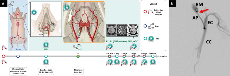
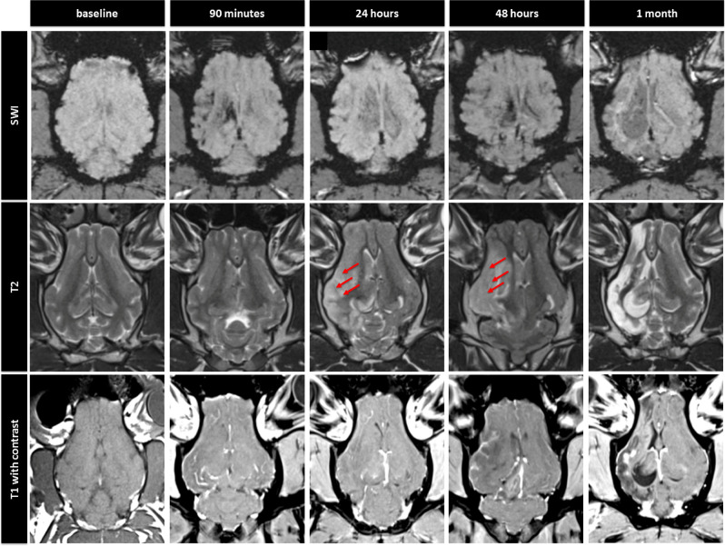
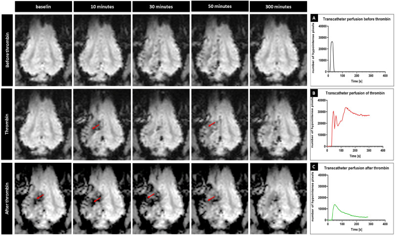
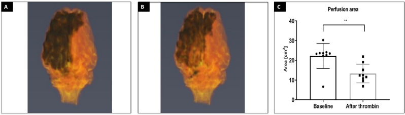
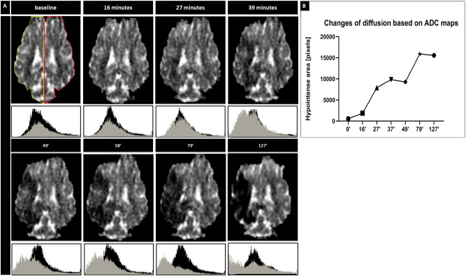
Modeling stroke in animals is essential for testing efficacy of new treatments; however, previous neuroprotective therapies, based on systemic delivery in rodents failed, exposing the need for model with improved clinical relevance. The purpose of this study was to develop endovascular approach for inducing ischemia in swine. To achieve that goal, we used intra-arterial administration of thrombin mixed with gadolinium and visualized the occlusion with real-time MRI. Placement of the microcatheter proximally to rete allowed trans-catheter perfusion of the ipsilateral hemisphere as visualized by contrast-enhanced perfusion MR scans. Dynamic T2*w MRI facilitated visualization of thrombin + Gd solution transiting through cerebral vasculature and persistent hyperintensities indicated occlusion. Area of trans-catheter perfusion dynamically quantified on representative slice before and after thrombin administration (22.20 ± 6.31 cm2 vs. 13.28 ± 4.71 cm2 respectively) indicated significantly reduced perfusion. ADC mapping showed evidence of ischemia as early as 27 min and follow-up T2w scans confirmed ischemic lesion (3.14 ± 1.41 cm2). Animals developed contralateral neurological deficits but were ambulatory. Our study has overcome long lasting challenge of inducing endovascular stroke model in pig. We were able to induce stroke using minimally invasive endovascular approach and observe in real-time formation of the thrombus, blockage of cerebral perfusion and eventually stroke lesion.


Conflict of interest statement

The authors declare no competing interests.

Figures

Figure 1
Figure 1
Experimental Outline. Timeline of experiments (A), Digital subtraction angiography with a catheter in APA (B; CC indicates common carotid artery, EC external carotid artery, AP ascending pharyngeal artery, RM rete mirabile, red arrow indicates placement of the microcatheter tip).
Figure 2
Figure 2
Longitudinal MR imaging of representative animal showing evolution of acute stroke. SWI, T2w (arrows indicate ischemic region) and T1 + Gd images at different time points.
Figure 3
Figure 3
Cerebral trans-catheter perfusion changes during induction of ischemia in representative animal. Perfusion changes over time at baseline, thrombin mixed with contrast agent injection and thrombin injection of representative animal (red arrows point to hypointense regions) with quantification (AC, respectively).
Figure 4
Figure 4
3D rendering of cerebral trans-catheter perfusion territory. 3D reconstruction of trans-catheter perfusion territory over time before (A) and after thrombin injection of representative animal (B) with quantification (C).
Figure 5
Figure 5
Diffusion changes after stroke induction. ADC over time with histograms for ipsilateral (grey) and contralateral (black) hemisphere (A). ROI for evaluation of changes in diffusion (A; yellow—ipsilateral and red—contralateral; X axis represents pixel intensity and Y axis represents number of pixels in that particular tone). ADC maps with quantification of brain area with abnormal ADC over time (B; apostrophes indicate minutes).
Figure 6
Figure 6
Histological evaluation of stroke tissue. BBB evaluation at 1 day, 1 week, and 3 months post stroke (A). Quantification of Intensity Mean Value (B). Hematoxylin and Eosin staining of the brain 3 months post stroke (C—contralateral hemisphere, D—ipsilateral hemisphere).

References

    1. Benjamin EJ, et al. Heart disease and stroke statistics-2018 Update: A report from the American Heart Association. Circulation. 2018;137:e67–e492. doi: 10.1161/CIR.0000000000000558. - DOI - PubMed
    1. National Institute of Neurological Disorders and Stroke rt-PA Stroke Study Group Tissue plasminogen activator for acute ischemic stroke. N. Engl. J. Med. 1995;333:1581–1587. doi: 10.1056/NEJM199512143332401. - DOI - PubMed
    1. Goyal M, et al. Endovascular thrombectomy after large-vessel ischaemic stroke: A meta-analysis of individual patient data from five randomised trials. Lancet. 2016;387:1723–1731. doi: 10.1016/S0140-6736(16)00163-X. - DOI - PubMed
    1. Yasuhara T, et al. Notch-induced rat and human bone marrow stromal cell grafts reduce ischemic cell loss and ameliorate behavioral deficits in chronic stroke animals. Stem Cells Dev. 2009;18:1501–1514. doi: 10.1089/scd.2009.0011. - DOI - PubMed
    1. Steinberg GK, et al. Two-year safety and clinical outcomes in chronic ischemic stroke patients after implantation of modified bone marrow-derived mesenchymal stem cells (SB623): A phase 1/2a study. J. Neurosurg. 2018 doi: 10.3171/2018.5.JNS173147. - DOI - PubMed

Publication types

MeSH terms