. 2020 Oct 14;10(1):17314.
doi: 10.1038/s41598-020-73997-y.

Nucleosomal embedding reshapes the dynamics of abasic sites

Affiliations

Nucleosomal embedding reshapes the dynamics of abasic sites

Emmanuelle Bignon et al. Sci Rep. .

Abstract

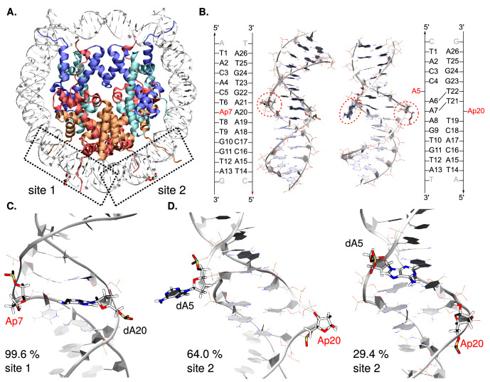
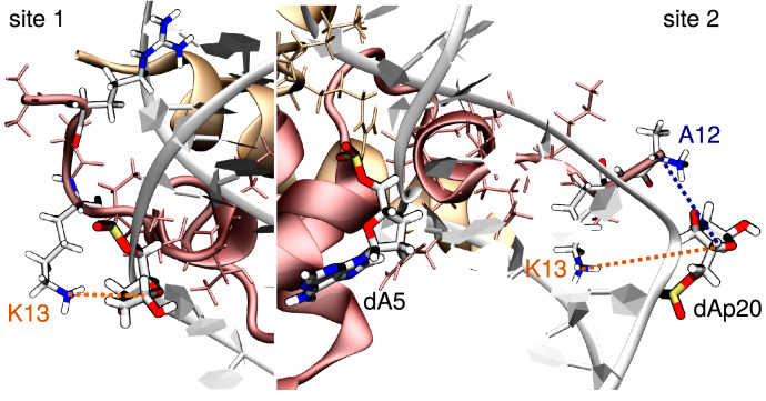
Apurinic/apyrimidinic (AP) sites are the most common DNA lesions, which benefit from a most efficient repair by the base excision pathway. The impact of losing a nucleobase on the conformation and dynamics of B-DNA is well characterized. Yet AP sites seem to present an entirely different chemistry in nucleosomal DNA, with lifetimes reduced up to 100-fold, and the much increased formation of covalent DNA-protein cross-links leading to strand breaks, refractory to repair. We report microsecond range, all-atom molecular dynamics simulations that capture the conformational dynamics of AP sites and their tetrahydrofuran analogs at two symmetrical positions within a nucleosome core particle, starting from a recent crystal structure. Different behaviours between the deoxyribo-based and tetrahydrofuran-type abasic sites are evidenced. The two solvent-exposed lesion sites present contrasted extrahelicities, revealing the crucial role of the position of a defect around the histone core. Our all-atom simulations also identify and quantify the frequency of several spontaneous, non-covalent interactions between AP and positively-charged residues from the histones H2A and H2B tails that prefigure DNA-protein cross-links. Such an in silico mapping of DNA-protein cross-links gives important insights for further experimental studies involving mutagenesis and truncation of histone tails to unravel mechanisms of DPCs formation.


Conflict of interest statement

The authors declare no competing interests.

Figures

Figure 1
Figure 1
(A) Chemical structure of an AP site, its THF analog, and DNA-protein cross-link (DPC) formed upon reaction between a lysine and an AP site. (B) Cartoon representation of the crystal structure of a nucleosome core particle harboring two symmetrical solvent-exposed tetrahydrofuran residues (sites 1 and 2; PDB ID 5JRG). Site 1 exhibits a classical B-DNA-like conformation, while site 2 adopts the so-called inchworm conformation following the exclusion of THF and of an adenine on the opposite strand. Biomolecule pictures were rendered with VMD 1.9.4a37 (https://www.ks.uiuc.edu/Research/vmd/),. All the figures were rearranged using Inkscape 0.91 (https://inkscape.org/).
Figure 2
Figure 2
(A) Structure of the THF-containing NCP (PDB ID 5JRG). Histones H2A, H2B, H3, and H4 appear in red, orange, blue, and cyan, respectively. The two symmetric lesion sites are depicted in licorice, site 1 on the left (B-DNA like conformation) and site 2 on the right (inchworm conformation). (B) Cartoon representation of the NCP 15-bp sections harboring the lesion sites, and their corresponding sequences. The damaged sites (here AP), as well as the in silico modeled dA5 for site 2, are encircled in red. Structural analyses were performed on these 15-bp sections only. Reference MD simulations of naked DNA were performed on the same sequences, with one extra base pair at each extremity to avoid edge effects. (C) and (D) Representative structures of the main clusters and their frequency of occurrence for site 1 (C) and site 2 (D) in AP-containing NCP. The lesion sites, the facing base for site 1 (dA20) and the ejected dA5 for site 2 are depicted in licorice. Conformations sampled for site 1 mostly belong to one dominant cluster (99.6% of the simulation time) while site 2 exhibits two main different conformations (64.0% and 29.4% of the simulation time). Pictures were rendered with VMD 1.9.4a37 (https://www.ks.uiuc.edu/Research/vmd/),.
Figure 3
Figure 3
Representation of the per-residue relative contribution to the 10 first principal components around site 1 (top) and site 2 (bottom), detailed for the three MD replicates with AP sites. Only DNA and the N-term tails of H2A and H2B are represented. The percentage of contribution of each residue ranges from 0% (blue) to 15% (red). Abasic sites, its facing orphan base, and the ejected dA5 at site 2 are depicted and encircled in red, magenta and brown, respectively. For a representation of the full NCP, see Figure S6. PCA pictures were rendered using open-source Pymol Version 1.8 (https://pymol.org).
Figure 4
Figure 4
Representation of the main interactions between the damaged site (here AP) and H2A K13 near site 1 (left) and H2A K13 and N-term A12 near site 2 (right). H2B appears in orange and H2A in red, with their tail residues in thin licorice. The abasic sites (dAp7 at site 1 and dAp20 at site 2), the ejected adenine at site 2 (dA5), and the interacting tail residues (K13, N-term A12, and R11) are depicted in thick licorice, colored by element. Pictures were rendered with VMD 1.9.4a37 (https://www.ks.uiuc.edu/Research/vmd/),.

References

    1. Greenberg MM. Abasic and oxidized abasic site reactivity in DNA: Enzyme inhibition, cross-linking, and nucleosome catalyzed reactions. Acc. Chem. Res. 2014;47:646–655. doi: 10.1021/ar400229d. - DOI - PMC - PubMed
    1. Wilson DM, Barsky D. The major human abasic endonuclease: Formation, consequences and repair of abasic lesions in DNA. Mutat. Res. DNA Repair. 2001;485:283–307. doi: 10.1016/S0921-8777(01)00063-5. - DOI - PubMed
    1. Drohat AC, Maiti A. Mechanisms for enzymatic cleavage of the n-glycosidic bond in DNA. Org. Biomol. Chem. 2014;12:8367–8378. doi: 10.1039/C4OB01063A. - DOI - PMC - PubMed
    1. Kitsera N, Rodriguez-Alvarez M, Emmert S, Carell T, Khobta A. Nucleotide excision repair of abasic DNA lesions. Nucleic Acids Res. 2019;47:8537–8547. doi: 10.1093/nar/gkz558. - DOI - PMC - PubMed
    1. Dutta S, Chowdhury G, Gates KS. Interstrand cross-links generated by abasic sites in duplex DNA. J. Am. Chem. Soc. 2007;129:1852–1853. doi: 10.1021/ja067294u. - DOI - PMC - PubMed

Publication types