. 2020 Oct 15;15(10):e0240227.
doi: 10.1371/journal.pone.0240227. eCollection 2020.

Auditory cortical activity elicited by infrared laser irradiation from the outer ear in Mongolian gerbils

Affiliations

Auditory cortical activity elicited by infrared laser irradiation from the outer ear in Mongolian gerbils

Yuta Tamai et al. PLoS One. .

Abstract

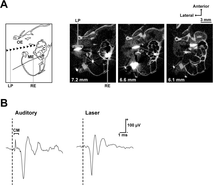
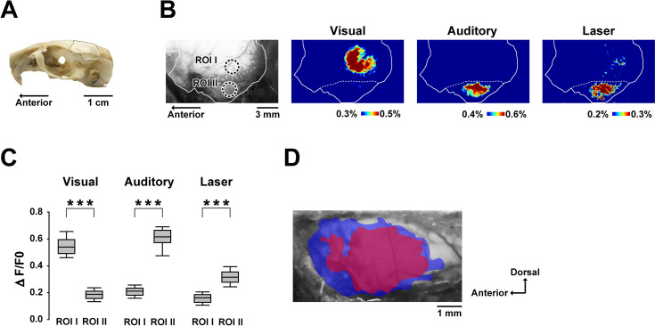
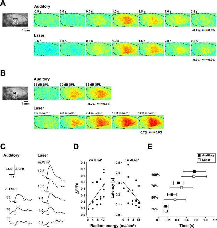
Infrared neural stimulation has been studied for its potential to replace an electrical stimulation of a cochlear implant. No studies, however, revealed how the technic reliably evoke auditory cortical activities. This research investigated the effects of cochlear laser stimulation from the outer ear on auditory cortex using brain imaging of activity-dependent changes in mitochondrial flavoprotein fluorescence signal. An optic fiber was inserted into the gerbil's ear canal to stimulate the lateral side of the cochlea with an infrared laser. Laser stimulation was found to activate the identified primary auditory cortex. In addition, the temporal profile of the laser-evoked responses was comparable to that of the auditory responses. Our results indicate that infrared laser irradiation from the outer ear has the capacity to evoke, and possibly manipulate, the neural activities of the auditory cortex and may substitute for the present cochlear implants in future.


Conflict of interest statement

The authors have declared that no competing interests exist.

Figures

Fig 1
Fig 1. Laser irradiation site and the cochlear responses to laser and auditory stimuli.
(A) Schematic of recording and stimulation configuration (the left-most) and microcomputed tomography images of a stimulated cochlea (horizontal section). The distance from the sagittal suture is stated on the bottom left of each image. The recording electrode (silver wire) and the laser path (tungsten wire) are shown as larger than their actual sizes due to metal artifacts in CT images. (B) Cochlear responses to auditory (100 μs single click with 70 dB peak-to-peak equivalent SPL) and laser (100 μs single pulses, 4.9 mJ/cm2) stimuli. A clear cochlear microphonic (CM) was observed only after the auditory stimulus. The vertical dashed lines show stimulus onset. Abbreviations: OE, outer ear; ME, middle ear; C, cochlea; LP, laser path; RE, recording electrode; CM, cochlear microphonic.
Fig 2
Fig 2. Cortical regions activated by visual, auditory and laser stimuli.
(A) Photograph showing the recording site (dashed line). (B) An original fluorescence image showing the ROI and pseudocolor images of ΔF/F0 of the left hemisphere. The solid line indicates the recording site and the dashed line indicates the ridge of the parietal bone. Each pseudocolor image is averaged over a period of 500 ms around the peak response time. (C) Amplitudes of visual-, auditory-, and laser-evoked responses for each ROI. The positions of ROI I and ROI II, depicted in (B), are centered over the peak responses corresponding to the visual and auditory stimuli, respectively. The box plots show the median and 25th and 75th percentiles, with whiskers at the 10th and 90th percentiles. ***P < 0.001 (Mann–Whitney U test with Bonferroni correction for multiple comparisons). (D) Positional relationship between the auditory- (blue) and laser-evoked (red) areas of the temporal region. The colored areas superimposed on the original fluorescence image show the cortical regions with a 75% maximum ΔF/F0 response. Data from 11 subjects were aligned based on the cerebral vascular pattern and were pooled.
Fig 3
Fig 3. Auditory- and laser-evoked flavoprotein responses in the temporal cortex.
(A) Original fluorescence image (left) and flavoprotein responses evoked by auditory (85 dB SPL) and laser (7.4 mJ/cm2) stimuli (right). The circular window shows the region of interest (ROI) centered on the maximum responses of all recordings. The recording areas are indicated with black lines. (B) Original fluorescence image and changes in intensity-dependent responses to auditory (55–85 dB SPL) and laser (0.5–12.8 mJ/cm2) stimuli. (C) Time-course of the fluorescence changes in the ROI following acoustic (55–85 dB SPL) and laser (0.5–12.8 mJ/cm2) stimuli. The vertical bars on the bottom represent the stimulus period. (D) Changes in response amplitude and response latency following infrared laser stimulation of various radiant energy (n = 6). (E) Latencies to the 25%, 50%, 75% and 100% maximum response to laser (7.4 mJ/cm2) and equivalent auditory stimuli (n = 12). Error bars indicate the standard deviation.

References

    1. Terry B, Kelt RE, Jeyakumar A. Delayed complications after cochlear implantation. JAMA otolaryngology—head & neck surgery. 2015;141(11):1012–7. Epub 2015/10/16. 10.1001/jamaoto.2015.2154 . - DOI - PubMed
    1. Friesen LM, Shannon RV, Baskent D, Wang X. Speech recognition in noise as a function of the number of spectral channels: comparison of acoustic hearing and cochlear implants. J Acoust Soc Am. 2001;110(2):1150–63. Epub 2001/08/25. 10.1121/1.1381538 . - DOI - PubMed
    1. Wells J, Kao C, Mariappan K, Albea J, Jansen ED, Konrad P, et al. Optical stimulation of neural tissue in vivo. Optics letters. 2005;30(5):504–6. Epub 2005/03/26. 10.1364/ol.30.000504 . - DOI - PubMed
    1. Wells J, Konrad P, Kao C, Jansen ED, Mahadevan-Jansen A. Pulsed laser versus electrical energy for peripheral nerve stimulation. J Neurosci Methods. 2007;163(2):326–37. Epub 2007/06/01. 10.1016/j.jneumeth.2007.03.016 - DOI - PMC - PubMed
    1. Wells J, Kao C, Konrad P, Milner T, Kim J, Mahadevan-Jansen A, et al. Biophysical mechanisms of transient optical stimulation of peripheral nerve. Biophys J. 2007;93(7):2567–80. Epub 2007/05/29. 10.1529/biophysj.107.104786 - DOI - PMC - PubMed

Publication types