. 2020 Oct 15;15(10):e0240333.
doi: 10.1371/journal.pone.0240333. eCollection 2020.

Fgf10-CRISPR mosaic mutants demonstrate the gene dose-related loss of the accessory lobe and decrease in the number of alveolar type 2 epithelial cells in mouse lung

Affiliations

Fgf10-CRISPR mosaic mutants demonstrate the gene dose-related loss of the accessory lobe and decrease in the number of alveolar type 2 epithelial cells in mouse lung

Munenori Habuta et al. PLoS One. .

Abstract

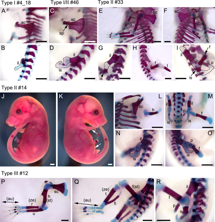
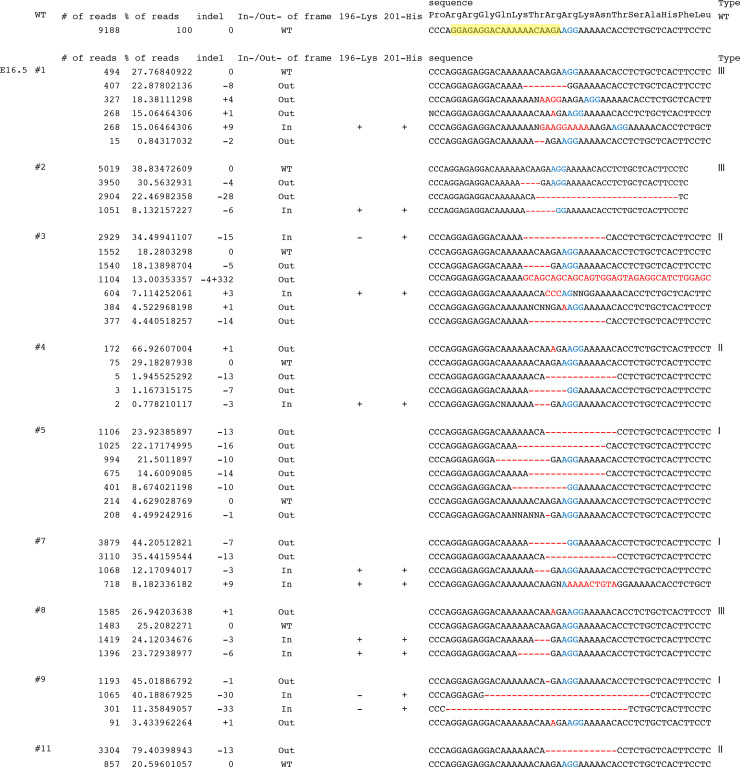
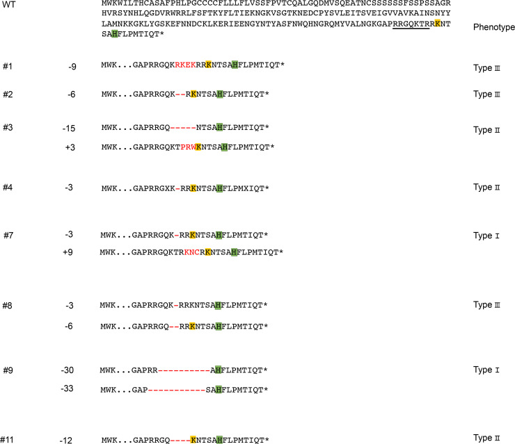
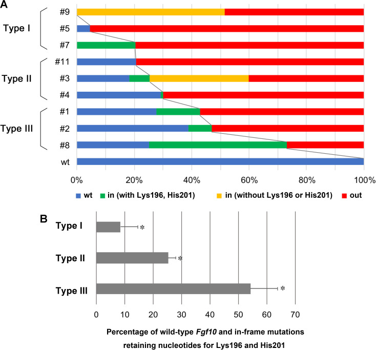
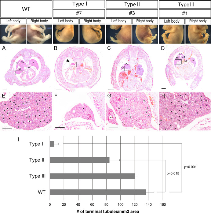
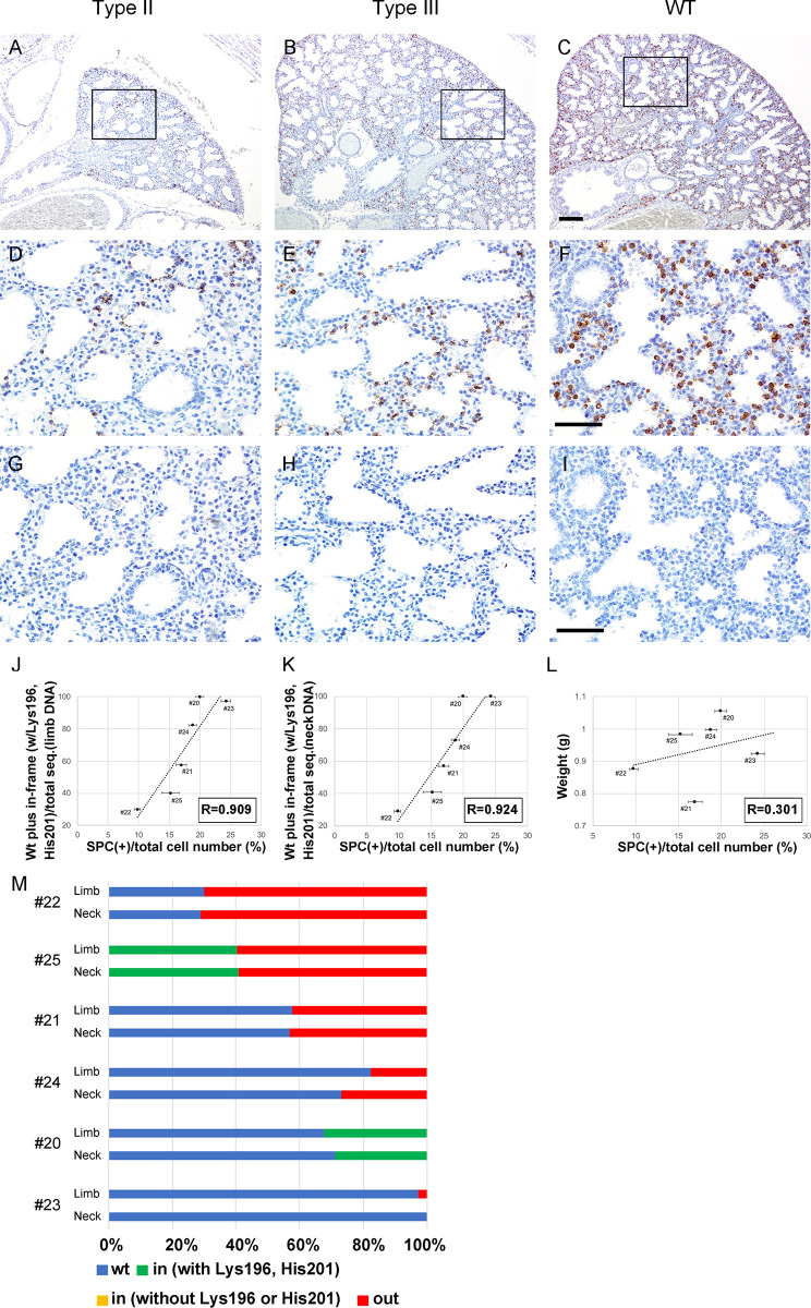
CRISPR/Cas9-mediated gene editing often generates founder generation (F0) mice that exhibit somatic mosaicism in the targeted gene(s). It has been known that Fibroblast growth factor 10 (Fgf10)-null mice exhibit limbless and lungless phenotypes, while intermediate limb phenotypes (variable defective limbs) are observed in the Fgf10-CRISPR F0 mice. However, how the lung phenotype in the Fgf10-mosaic mutants is related to the limb phenotype and genotype has not been investigated. In this study, we examined variable lung phenotypes in the Fgf10-targeted F0 mice to determine if the lung phenotype was correlated with percentage of functional Fgf10 genotypes. Firstly, according to a previous report, Fgf10-CRISPR F0 embryos on embryonic day 16.5 (E16.5) were classified into three types: type I, no limb; type II, limb defect; and type III, normal limbs. Cartilage and bone staining showed that limb truncations were observed in the girdle, (type I), stylopodial, or zeugopodial region (type II). Deep sequencing of the Fgf10-mutant genomes revealed that the mean proportion of codons that encode putative functional FGF10 was 8.3 ± 6.2% in type I, 25.3 ± 2.7% in type II, and 54.3 ± 9.5% in type III (mean ± standard error of the mean) mutants at E16.5. Histological studies showed that almost all lung lobes were absent in type I embryos. The accessory lung lobe was often absent in type II embryos with other lobes dysplastic. All lung lobes formed in type III embryos. The number of terminal tubules was significantly lower in type I and II embryos, but unchanged in type III embryos. To identify alveolar type 2 epithelial (AECII) cells, known to be reduced in the Fgf10-heterozygous mutant, immunostaining using anti-surfactant protein C (SPC) antibody was performed: In the E18.5 lungs, the number of AECII was correlated to the percentage of functional Fgf10 genotypes. These data suggest the Fgf10 gene dose-related loss of the accessory lobe and decrease in the number of alveolar type 2 epithelial cells in mouse lung. Since dysfunction of AECII cells has been implicated in the pathogenesis of parenchymal lung diseases, the Fgf10-CRISPR F0 mouse would present an ideal experimental system to explore it.


Conflict of interest statement

Supported by an academic grant from Pfizer Japan, Inc. This does not alter our adherence to PLOS ONE policies on sharing data and materials.

Figures

Fig 1
Fig 1. Cartilage and bone staining to reveal limb skeletal structures.
A-I, L-R, left or right lateral views of limb and girdle regions (at E18.5) are shown. Cartilage is stained with Alcian blue and bone is stained with Alizarin red. A, B, type I #4_18 embryo. C, D, type I/II #46 embryo. E-I, type II #33 embryo. L-O, type II #14 embryo. P-R, type III #12 embryo. J, K, Lateral views of type II #14 embryo are shown. Arrows in (K) show truncated limbs. au, autopod; f, femur; fi, fibula; h, humerus; il, ilium; is, ischium; p, pubis; r, radius; sc, scapula; sp, spine; st, stylopod t, tibia; u, ulna; ze, zeugopod. Scale bars: 1 mm.
Fig 2
Fig 2. Genomic analysis of the Fgf10-CRISPR F0 embryos at E16.5 as revealed by deep sequencing.
The target nucleotide sequence is highlighted in yellow. Proto-spacer adjacent motif sequence is shown in blue. Insertion and deletion sequences are shown in red.
Fig 3
Fig 3. Deduced amino acids for in-frame mutations after deep sequencing.
Wild type (WT) sequences are shown on the top. The embryo (E16.5) number (#), nucleotide number of small insertion (+) or deletion (-) (Indels) are shown on the left. Classified “types” are shown on the right. Amino acids corresponding to the guide RNA sequence are underlined. Deleted and altered amino acids are indicated in red.
Fig 4
Fig 4. Schematic representation of genomic analysis of Fgf10-CRISPR F0 embryos at E16.5 by deep sequencing.
A, percentage of total reads for the Fgf10 crispants. wt, wild type Fgf10 target nucleotide sequence; in, in-frame mutations by small insertion or deletion; out, frameshift mutations. Among in-frame mutations, the percentage of those that preserve the codons for Lys196 and His201 is shown in green, while the percentage that eliminate either of them or both is shown in yellow. B, percentage of wild type and in-frame mutations in which nucleotides for both Lys196 and His201 are retained: 8.3 ± 6.2% for type I, 25.3 ± 2.7% for type II, and 54.3 ± 9.5% for type III. Data are presented as means ± SEM. *p<0.01 (p = 0.008).
Fig 5
Fig 5. Limb phenotypes and lung histology of Fgf10-CRISPR F0 embryos at E16.5.
Representative embryos are shown for wild type (WT), type I (embryo #7), type II (#3), and type III (#1). A–D, transverse section of the embryonic chest region. E–H, close-up of the embryonic lung (boxed area) shown in (A–D), respectively. Asterisks show putative terminal tubules in the lung. I, the number of lung terminal tubules per unit area. In type I and type II embryos, there is a significant decrease in the number compared with that of wild type. Data are presented as means ± SEM. Source data are available in Table 3. Ac, accessory lobe; Ca, caudal lobe; Cr, cranial lobe; L, left lobe; Mi, middle lobe. Scale bars: 500 μm in (A–D), and 100 μm in (E–H).
Fig 6
Fig 6. Immunohistochemistry of the lung in type II and type III Fgf10-crispants and wild type embryos at E18.5.
Representative data are shown. Nuclei are stained with Hematoxylin. A-C, Localization of Surfactant protein C (SPC) (alveolar type 2 epithelial cells) is indicated by brown staining. D-F, close-up of the boxed area shown in (A–C), respectively. G-I, negative control, using normal rabbit IgG instead of anti-SPC antibody. Scale bars: 100 μm in (A-C); 50 μm in (D-I). J, K, the number of SPC-positive cells in type III embryos is correlated to the percentage of functional Fgf10 genotypes in limb (J) or neck DNA (K). L, the number of SPC-positive cells in type III embryos is not to correlated to their weight (K). Source data for (J-L) are available in Table 1. M, Schematic drawings to show percentage of total reads for the type III Fgf10 crispants. wt, wild type Fgf10 target nucleotide sequence; in, in-frame mutations by small insertion or deletion; out, out-of-frame mutations. Among in-frame mutations, the percentage of those that preserve the codons for Lys196 and His201 is shown in green, while the percentage that eliminate either of them is shown in yellow.

Similar articles

Cited by

References

    1. Doudna JA, Charpentier E. Genome editing. The new frontier of genome engineering with CRISPR-Cas9. Science. 2014;346(6213):1258096 10.1126/science.1258096 - DOI - PubMed
    1. Hsu PD, Lander ES, Zhang F. Development and applications of CRISPR-Cas9 for genome engineering. Cell. 2014;157(6):1262–78. 10.1016/j.cell.2014.05.010 - DOI - PMC - PubMed
    1. Cornu TI, Mussolino C, Cathomen T. Refining strategies to translate genome editing to the clinic. Nat Med. 2017;23(4):415–23. 10.1038/nm.4313 - DOI - PubMed
    1. Sung YH, Kim JM, Kim HT, Lee J, Jeon J, Jin Y, et al. Highly efficient gene knockout in mice and zebrafish with RNA-guided endonucleases. Genome Res. 2014;24(1):125–31. 10.1101/gr.163394.113 - DOI - PMC - PubMed
    1. Yang H, Wang H, Shivalila CS, Cheng AW, Shi L, Jaenisch R. One-step generation of mice carrying reporter and conditional alleles by CRISPR/Cas-mediated genome engineering. Cell. 2013;154(6):1370–9. 10.1016/j.cell.2013.08.022 - DOI - PMC - PubMed

Publication types

MeSH terms

Substances

LinkOut - more resources