

Characterization of hepatic macrophages and evaluation of inflammatory response in heme oxygenase-1 deficient mice exposed to scAAV9 vectors
- PMID: 33057437
- PMCID: PMC7561190
- DOI: 10.1371/journal.pone.0240691
Characterization of hepatic macrophages and evaluation of inflammatory response in heme oxygenase-1 deficient mice exposed to scAAV9 vectors
Abstract
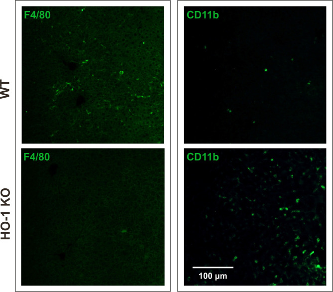
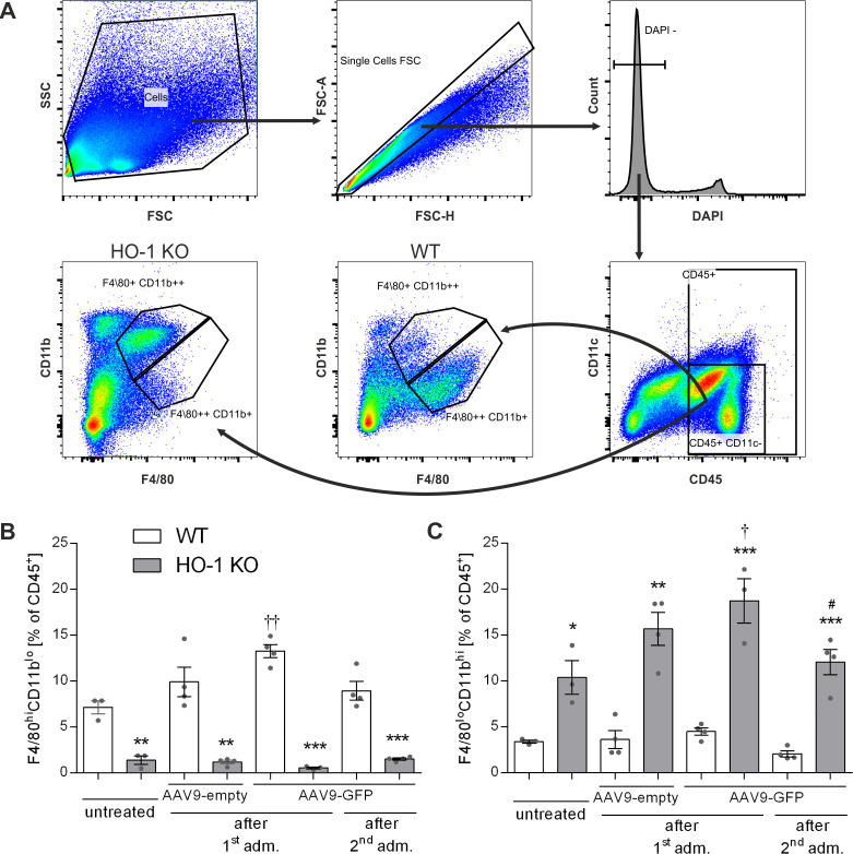
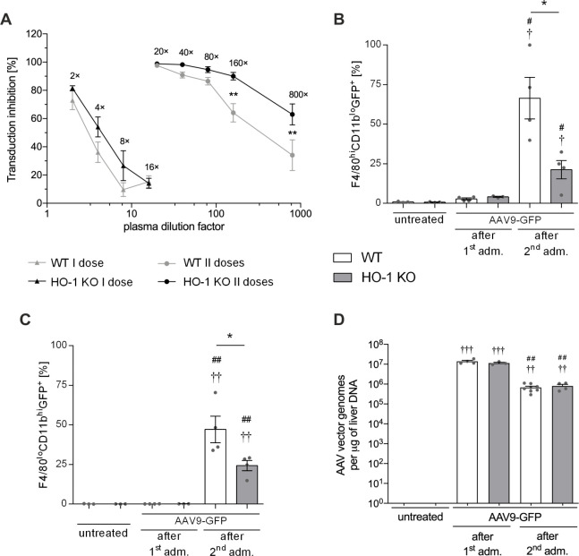
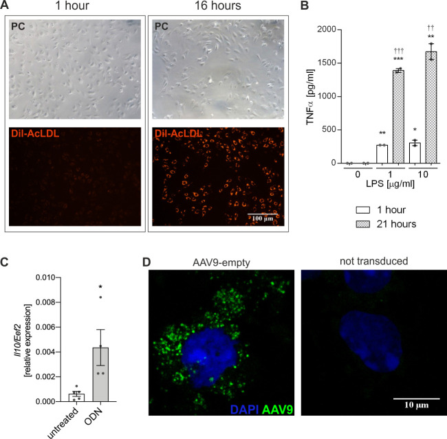
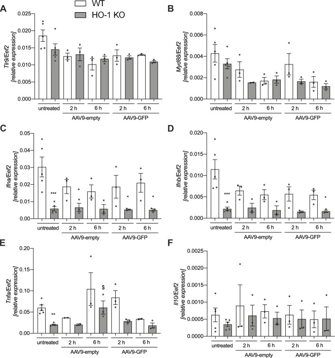
Adeno-associated viral (AAV) vectors are characterised by low immunogenicity, although humoral and cellular responses may be triggered upon infection. Following systemic administration high levels of vector particles accumulate within the liver. Kupffer cells (KCs) are liver resident macrophages and an important part of the liver innate immune system. Decreased functional activity of KCs can contribute to exaggerated inflammatory response upon antigen exposure. Heme oxygenase-1 (HO-1) deficiency is associated with considerably reduced numbers of KCs. In this study we aimed to investigate the inflammatory responses in liver and to characterise two populations of hepatic macrophages in adult wild type (WT) and HO-1 knockout (KO) mice following systemic administration of one or two doses (separated by 3 months) of self-complementary (sc)AAV9 vectors. At steady state, the livers of HO-1 KO mice contained significantly higher numbers of monocyte-derived macrophages (MDMs), but significantly less KCs than their WT littermates. Three days after re-administration of scAAV9 we observed increased mRNA level of monocyte chemoattractant protein-1 (Mcp-1) in the livers of both WT and HO-1 KO mice, but the protein level and the macrophage infiltration were not affected. Three days after the 1st and 3 days after the 2nd vector dose the numbers of AAV genomes in the liver were comparable between both genotypes indicating similar transduction efficiency, but the percentage of transgene-expressing MDMs and KCs was higher in WT than in HO-1 KO mice. In the primary culture, KCs were able to internalize AAV9 particles without induction of TLR9-mediated immune responses, but no transgene expression was observed. In conclusion, in vivo and in vitro cultured KCs have different susceptibility to scAAV9 vectors. Regardless of the presence or absence of HO-1 and initial numbers of KCs in the liver, scAAV9 exhibits a low potential to stimulate inflammatory response at the analysed time points.
Conflict of interest statement
The authors have declared that no competing interests exist.
Figures

Similar articles
-
The genome of self-complementary adeno-associated viral vectors increases Toll-like receptor 9-dependent innate immune responses in the liver.Blood. 2011 Jun 16;117(24):6459-68. doi: 10.1182/blood-2010-10-314518. Epub 2011 Apr 7. Blood. 2011. PMID: 21474674 Free PMC article.
-
Viral interleukin-10 gene transfer prevents liver ischemia-reperfusion injury: Toll-like receptor-4 and heme oxygenase-1 signaling in innate and adaptive immunity.Hum Gene Ther. 2007 Apr;18(4):355-66. doi: 10.1089/hum.2007.181. Hum Gene Ther. 2007. PMID: 17439357
-
Liver X receptors regulate hepatic F4/80 + CD11b+ Kupffer cells/macrophages and innate immune responses in mice.Sci Rep. 2018 Jun 18;8(1):9281. doi: 10.1038/s41598-018-27615-7. Sci Rep. 2018. PMID: 29915246 Free PMC article.
-
Anti-inflammatory pathways and alcoholic liver disease: role of an adiponectin/interleukin-10/heme oxygenase-1 pathway.World J Gastroenterol. 2010 Mar 21;16(11):1330-6. doi: 10.3748/wjg.v16.i11.1330. World J Gastroenterol. 2010. PMID: 20238399 Free PMC article. Review.
-
Heme oxygenase-1 and anti-inflammatory M2 macrophages.Arch Biochem Biophys. 2014 Dec 15;564:83-8. doi: 10.1016/j.abb.2014.09.005. Epub 2014 Sep 18. Arch Biochem Biophys. 2014. PMID: 25241054 Review.
Cited by
-
Single-cell transcriptomics reveals subtype-specific molecular profiles in Nrf2-deficient macrophages from murine atherosclerotic aortas.Front Immunol. 2023 Oct 27;14:1249379. doi: 10.3389/fimmu.2023.1249379. eCollection 2023. Front Immunol. 2023. PMID: 37965327 Free PMC article.
-
Gene therapy prevents onset of mitochondrial cardiomyopathy in neonatal mice with Ndufs6 deficiency.Cell Death Discov. 2025 May 22;11(1):249. doi: 10.1038/s41420-025-02524-7. Cell Death Discov. 2025. PMID: 40399258 Free PMC article.
References
-
- McCarty DM. Self-complementary AAV vectors; advances and applications. Mol Ther J Am Soc Gene Ther. 2008. October;16(10):1648–56. - PubMed
-
- Zincarelli C, Soltys S, Rengo G, Rabinowitz JE. Analysis of AAV serotypes 1–9 mediated gene expression and tropism in mice after systemic injection. Mol Ther J Am Soc Gene Ther. 2008. June;16(6):1073–80. - PubMed
Publication types
MeSH terms
Substances
LinkOut - more resources
Full Text Sources
Molecular Biology Databases
Research Materials
Miscellaneous
