Review
. 2020 Oct 1;20(10):17.
doi: 10.1167/jov.20.10.17.

What do radiologists look for? Advances and limitations of perceptual learning in radiologic search

Affiliations
Review

What do radiologists look for? Advances and limitations of perceptual learning in radiologic search

Robert G Alexander et al. J Vis. .

Abstract

Supported by guidance from training during residency programs, radiologists learn clinically relevant visual features by viewing thousands of medical images. Yet the precise visual features that expert radiologists use in their clinical practice remain unknown. Identifying such features would allow the development of perceptual learning training methods targeted to the optimization of radiology training and the reduction of medical error. Here we review attempts to bridge current gaps in understanding with a focus on computational saliency models that characterize and predict gaze behavior in radiologists. There have been great strides toward the accurate prediction of relevant medical information within images, thereby facilitating the development of novel computer-aided detection and diagnostic tools. In some cases, computational models have achieved equivalent sensitivity to that of radiologists, suggesting that we may be close to identifying the underlying visual representations that radiologists use. However, because the relevant bottom-up features vary across task context and imaging modalities, it will also be necessary to identify relevant top-down factors before perceptual expertise in radiology can be fully understood. Progress along these dimensions will improve the tools available for educating new generations of radiologists, and aid in the detection of medically relevant information, ultimately improving patient health.


Figures
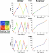
Figure 1.
Figure 1.
Description of 3D scan paths from Drew et al. (2013), who recorded eye position in each quadrant (left panel) as observers scrolled through CT scans in depth. Color indicates the quadrant of the image the radiologist was looking at during a given time in the trial. “Depth” on the y-axis refers to the 2D orthogonal slice of the scan currently viewed. In this study, radiologists looking for nodules on chest CTs could be characterized into two groups based on their search strategies. “Drillers,” such as the radiologist whose data appear in the middle column, tend to look within a single region of an image while quickly scrolling back and forth in depth through stacks of images. “Scanners,” such as the radiologist whose data appear in the right column, scroll more slowly in depth, and typically do not return to depths that they have already viewed. Scanners make more frequent eye movements to different spatial locations on the image, exploring the current 2D slice in greater detail. Note that although scanners spend more time than drillers making saccades per slice, neither scanners nor drillers visit all four quadrants of the image on every slice. Thus some regions of some slices may never be viewed foveally by either group. (Reprinted from Drew et al., 2013).
Figure 2.
Figure 2.
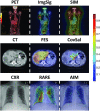
Examples of saliency models applied to PET, CT, and CXR. Saliency maps are represented as heat maps, with color indicating the saliency at that location: red is more salient than blue. The left column displays representative images. The middle column shows examples of saliency maps that accurately highlighted the regions of interest in the images. The right column shows examples of saliency maps that highlighted task-irrelevant regions in the images. Models with accurate predictions may provide insight into the features that radiologists use to view images, and models with inaccurate predictions may help narrow the list of potential features that need to be assessed. Image signature (ImgSig; Hou, Harel, & Koch, 2012), fast and efficient saliency (FES; Tavakoli, Rahtu, & Heikkilä, 2011), and RARE (Riche et al., 2013) were top-ranked models for PET, CT, and CXR. (Reprinted from Wen et al., 2017). SIM, Saliency by induction mechanisms; CovSal, Covariance saliency; AIM, Attention based on information maximization.

References

    1. Alexander R. G., Nahvi R. J., & Zelinsky G. J. (2019). Specifying the precision of guiding features for visual search. Journal of Experimental Psychology: Human Perception & Performance, 45(9), 1248–1264, doi:10.1037/xhp0000668. - DOI - PMC - PubMed
    1. Alexander R. G., Schmidt J., & Zelinsky G. J. (2014). Are summary statistics enough? Evidence for the importance of shape in guiding visual search. Visual Cognition, 22(3–4), 595–609, doi:10.1080/13506285.2014.890989. - DOI - PMC - PubMed
    1. Alexander R. G., & Zelinsky G. J. (2011). Visual similarity effects in categorical search. Journal of Vision, 11(8):9, 1–15, doi:10.1167/11.8.9. - DOI - PMC - PubMed
    1. Alzubaidi M., Balasubramanian V., Patel A., Panchanathan S., & Black Jr J. A. (2010, March). What catches a radiologist's eye? A comprehensive comparison of feature types for saliency prediction. In Medical Imaging 2010: Computer-Aided Diagnosis. International Society for Optics and Photonics, 7624, 76240W, doi:10.1016/j.visres.2011.12.004. - DOI
    1. Alexander R. G., & Zelinsky G. J. (2018). Occluded information is restored at preview but not during visual search. Journal of Vision, 11(4), 1–16, doi:10.1167/18.11.4. - DOI - PMC - PubMed

Publication types