Review
. 2020 Oct 14;115(6):63.
doi: 10.1007/s00395-020-00825-9.

Targeting myocardial ischaemic injury in the absence of reperfusion

Affiliations
Review

Targeting myocardial ischaemic injury in the absence of reperfusion

M V Basalay et al. Basic Res Cardiol. .

Abstract

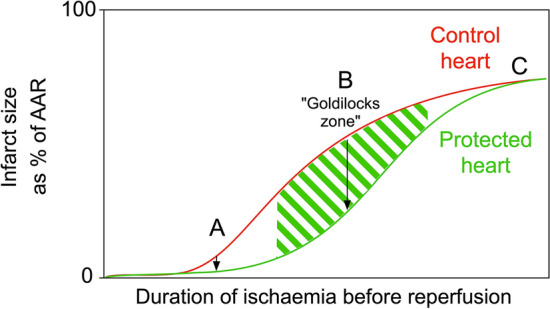
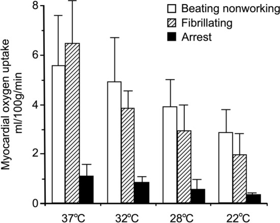
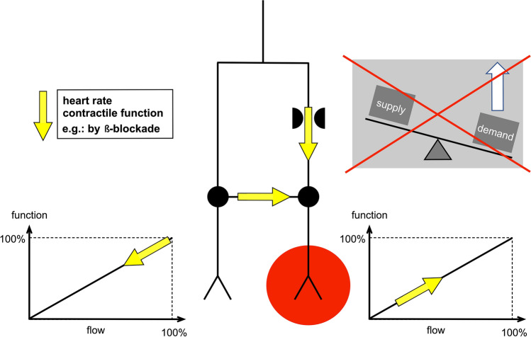
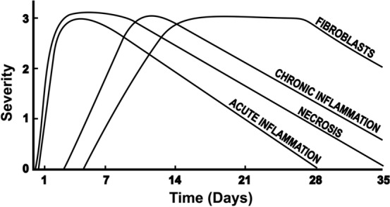
Sudden myocardial ischaemia causes an acute coronary syndrome. In the case of ST-elevation myocardial infarction (STEMI), this is usually caused by the acute rupture of atherosclerotic plaque and obstruction of a coronary artery. Timely restoration of blood flow can reduce infarct size, but ischaemic regions of myocardium remain in up to two-thirds of patients due to microvascular obstruction (MVO). Experimentally, cardioprotective strategies can limit infarct size, but these are primarily intended to target reperfusion injury. Here, we address the question of whether it is possible to specifically prevent ischaemic injury, for example in models of chronic coronary artery occlusion. Two main types of intervention are identified: those that preserve ATP levels by reducing myocardial oxygen consumption, (e.g. hypothermia; cardiac unloading; a reduction in heart rate or contractility; or ischaemic preconditioning), and those that increase myocardial oxygen/blood supply (e.g. collateral vessel dilation). An important consideration in these studies is the method used to assess infarct size, which is not straightforward in the absence of reperfusion. After several hours, most of the ischaemic area is likely to become infarcted, unless it is supplied by pre-formed collateral vessels. Therefore, therapies that stimulate the formation of new collaterals can potentially limit injury during subsequent exposure to ischaemia. After a prolonged period of ischaemia, the heart undergoes a remodelling process. Interventions, such as those targeting inflammation, may prevent adverse remodelling. Finally, harnessing of the endogenous process of myocardial regeneration has the potential to restore cardiomyocytes lost during infarction.

Keywords: Animals; Cardioprotection; Heart; Infarction; Ischaemia; Regeneration; Remodelling.


Conflict of interest statement

The authors declare that they have no conflict of interest.

Figures

Fig. 1
Fig. 1
The potential for cardioprotective strategies to limit myocardial infarction depends on the duration of ischaemia prior to reperfusion. If the duration of ischaemia before reperfusion is very short (a), then even if the relative reduction in infarct size is significant, the absolute reduction in infarct size will be very small. If the duration of ischaemia is excessively long (c), cardioprotective strategies will be ineffective. After intermediate durations of ischaemia followed by reperfusion (the “goldilocks” zone) (b), protection is maximal. Note that in this example, infarct size (as a percentage of ischaemic area at risk or AAR) is measured at a point after reperfusion, and accounts for cell death occurring during both ischaemia and reperfusion
Fig. 2
Fig. 2
a Infarct size expressed as a percentage of area at risk, vs time after coronary occlusion in rabbits, rats, pigs, dogs, cats and guinea pigs. Infarcts develop rapidly and completely in rats, rabbits and pigs. Redrawn from Schaper W et al. Prog Cardiovasc Dis. 1988; 31:57–77, with permission from Elsevier. b Collateral flow in different species, as a percentage of flow in the ischaemic vs non-ischaemic myocardium (mean ± SEM). n = number of hearts examined. Redrawn from Maxwell MP et al., Cardiovasc Res. 1987; 21(10):737–746, with permission from Oxford University Press
Fig. 3
Fig. 3
Myocardial oxygen uptake during cardiopulmonary bypass at different temperatures ranging from 37 to 22 °C. Redrawn from Buckberg GD. J Thorac Cardiovasc Surg. 1991; 102:895–903, with permission from Elsevier
Fig. 4
Fig. 4
Redistribution of coronary blood flow between ischaemic and non-ischaemic myocardium by beta-blockade. Reprinted from Heusch G. Am J Physiol Heart Circ Physiol. 2019; 316:H1439-H1446, with permission from G Heusch
Fig. 5
Fig. 5
Coronary collateral vessels can allow residual blood flow towards an ischaemic region. In what as known as “coronary steal phenomenon”, an increase in blood flow in a normal region of the myocardium can results in a decrease in blood flow to a partially ischaemic region (here indicated distal to an atheromatous plaque). Redrawn from Ritter et al., Rang & Dale's Pharmacology 8E, 2015:261, with permission from Elsevier
Fig. 6
Fig. 6
Course of time for histopathologic changes in myocardial infarction in man. Ordinate indicates relative severity of histopathologic changes. Redrawn from Fishbein MC et al. Chest 1978; 73:843–849, with permission from Elsevier

References

    1. Achour H, Boccalandro F, Felli P, Amirian J, Uthman M, Buja M, Smalling RW. Mechanical left ventricular unloading prior to reperfusion reduces infarct size in a canine infarction model. Catheter Cardiovasc Interv. 2005;64:182–192. doi: 10.1002/ccd.20271. - DOI - PubMed
    1. Ahn KT, Bin SY, Choe YH, Yang JH, Hahn JY, Choi JH, Choi SH, Chang SA, Lee SC, Lee SH, Oh JK, Gwon HC. Impact of transmural necrosis on left ventricular remodeling and clinical outcomes in patients undergoing primary percutaneous coronary intervention for ST-segment elevation myocardial infarction. Int J Cardiovasc Imaging. 2013;29:835–842. doi: 10.1007/s10554-012-0155-9. - DOI - PubMed
    1. Amanakis G, Kleinbongard P, Heusch G, Skyschally A. Attenuation of ST-segment elevation after ischemic conditioning maneuvers reflects cardioprotection online. Basic Res Cardiol. 2019;114:22. doi: 10.1007/s00395-019-0732-3. - DOI - PubMed
    1. Arcari L, Cimino S, De Luca L, Francone M, Galea N, Reali M, Carbone I, Iacoboni C, Agati L. Impact of heart rate on myocardial salvage in timely reperfused patients with ST-segment elevation myocardial infarction: new insights from cardiovascular magnetic resonance. PLoS ONE. 2015;10:e0145495. doi: 10.1371/journal.pone.0145495. - DOI - PMC - PubMed
    1. Badeer H. Effect of hypothermia on oxygen consumption and energy utilization of heart. Circ Res. 1956;4:523–526. doi: 10.1161/01.res.4.5.523. - DOI - PubMed

Publication types

MeSH terms

LinkOut - more resources