

Relation of Birthweight and Ovarian and Uterine Size Prior to Menarche
- PMID: 33058070
- PMCID: PMC8076108
- DOI: 10.1007/s43032-020-00351-y
Relation of Birthweight and Ovarian and Uterine Size Prior to Menarche
Abstract
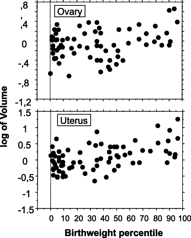
During pregnancy, supply of nutrients and exposure of the mother to environmental factors can influence fetus phenotype, possibly modifying growth of fetal tissues and organs. Few studies inconsistently reported that fetuses exposed to an insufficient energy supply, as those born small for gestational age, may have a reduced volume of uterus and ovaries. A retrospective analysis was performed on ultrasound data performed between 2012 and 2018 in 69 young premenarchal girls, 5 to 9 years of age, attending our endocrine-gynecologic clinic for a suspect of early puberty. Length of pregnancy and birthweight was also retrieved. When corrected for age, and presence of ovarian follicles, ovarian volume was positively (R2 = 0.210; p = 0.001) related to percentiles of birthweight (beta coefficient 0.012; 95% CI, 0.002-0.021). Similarly, uterine volume was positively (R2 = 0.237; p = 0.005) related to percentiles of birthweight (beta coefficient 0.067; 95% CI, 0.021-0.114). Ovarian (p = 0.034) and uterine (p = 0.014) volume was higher in the upper 3rd distribution of birthweight percentiles. In conclusion, development of ovarian and uterine volume increases progressively with the increase of birthweight percentiles. The data indicate an association between birthweight and the volume of uterus and ovary at 5-9 years of age.
Keywords: Birthweight; Fetal growth; Ovary; Reproduction; Small for gestational age; Uterus.
Conflict of interest statement
The authors declare that they have no competing interests.
Figures
References
-
- Ghirri P, Bernardini M, Vuerich M, Cuttano AM, Coccoli L, Merusi I, Ciulli C, D’Accavio L, Bottone U, Boldrini A. Adrenarche, pubertal development, age at menarche and final height of full-term, born small for gestational age (SGA) girls. Gynecol Endocrinol. 2001;15:91–97. - PubMed
-
- Ibáñez L, de Zegher F. Puberty after prenatal growth restraint. Horm Res. 2006;65(Suppl 3):112–115. - PubMed
Publication types
MeSH terms
LinkOut - more resources
Full Text Sources
Medical
