

Polyclonal B-cell lymphocytosis in English bulldogs
- PMID: 33058280
- PMCID: PMC7694837
- DOI: 10.1111/jvim.15913
Polyclonal B-cell lymphocytosis in English bulldogs
Abstract
Background: English bulldogs disproportionally develop an expansion of small B-cells, which has been interpreted as B-cell chronic lymphocytic leukemia (BCLL). However, clonality testing in these cases has often not been supportive of neoplasia.
Hypothesis: English bulldogs have a syndrome of nonneoplastic B-cell expansion.
Animals: Eighty-four English bulldogs with small-sized CD21+ B-cell lymphocytosis in the blood as determined by flow cytometry.
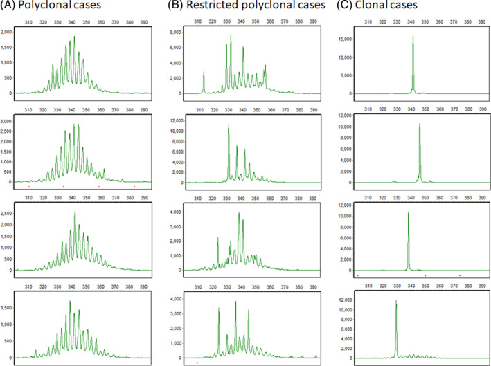
Methods: This is a retrospective study. We characterized this syndrome by assessing B-cell clonality, clinical presentation, flow cytometric features, and immunoglobulin gammopathy patterns. We identified 84 cases with CD21+ lymphocytosis among 195 English bulldogs with blood samples submitted to the Colorado State University-Clinical Immunology laboratory for immunophenotyping between 2010 and 2019. Flow cytometry features were compared to normal B-cells and BCLL cases. PCR for antigen receptor rearrangements (PARR) by multiple immunoglobulin primers was performed to assess B-cell clonality. A subset of cases with gammopathy were examined by protein electrophoresis, immunofixation, and immunoglobulin subclass ELISA quantification.
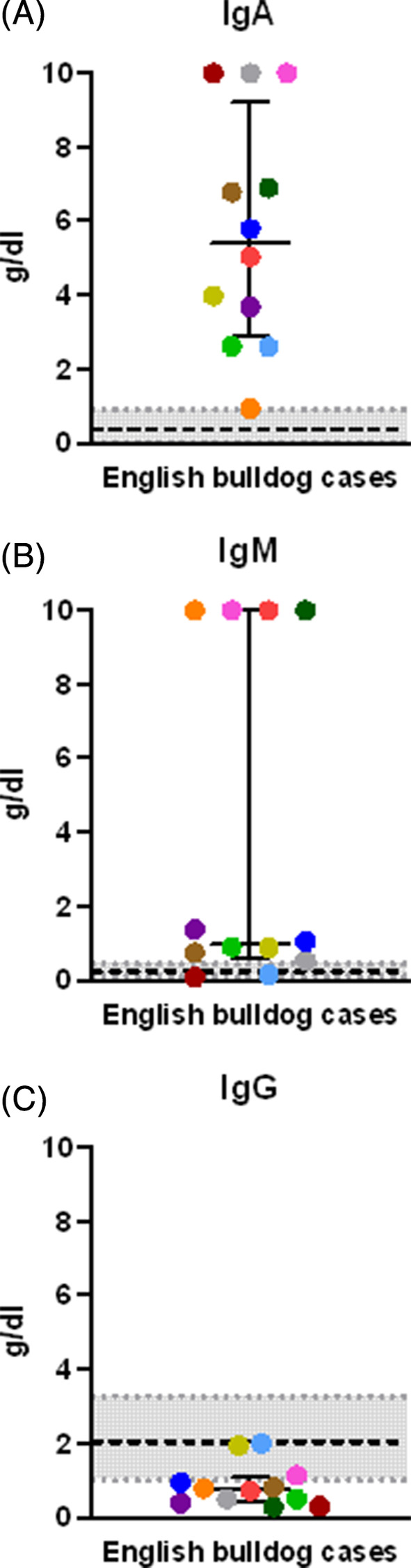
Results: Seventy percent (58/83) of cases had polyclonal or restricted polyclonal immunoglobulin gene rearrangements, suggesting nonmalignant B-cell expansion. The median age of all dogs in the study was 6.8 years and 74% were male. The median (range) lymphocyte count was 22 400/μL (2000-384 400/μL) and B-cells had low expression of class II MHC and CD25. Splenomegaly or splenic masses were detected in 57% (26/46) of cases and lymphadenopathy in 11% (7/61). Seventy-one percent (52/73) of cases had hyperglobulinemia and 77% (23/30) with globulin characterization had IgA ± IgM polyclonal or restricted polyclonal gammopathy patterns.
Conclusions and clinical importance: Polyclonal B-cell lymphocytosis in English bulldogs is characterized by low B-cell class II MHC and CD25 expression, splenomegaly and hyperglobulinemia consisting of increased IgA ± IgM. We hypothesize that this syndrome has a genetic basis.
Keywords: IgA gammopathy; PCR for antigen receptor rearrangements; canine; clinical pathology; clonality; flow cytometry; hyperglobulinemia.
© 2020 The Authors. Journal of Veterinary Internal Medicine published by Wiley Periodicals LLC on behalf of American College of Veterinary Internal Medicine.
Conflict of interest statement
Authors declare no conflict of interest.
Figures


Similar articles
-
Clinical outcome and prognostic factors in dogs with B-cell chronic lymphocytic leukemia: A retrospective study.J Vet Intern Med. 2021 Jul;35(4):1918-1928. doi: 10.1111/jvim.16160. Epub 2021 May 17. J Vet Intern Med. 2021. PMID: 33998726 Free PMC article.
-
Antibody tests for identification of current and past infection with SARS-CoV-2.Cochrane Database Syst Rev. 2022 Nov 17;11(11):CD013652. doi: 10.1002/14651858.CD013652.pub2. Cochrane Database Syst Rev. 2022. PMID: 36394900 Free PMC article.
-
Signs and symptoms to determine if a patient presenting in primary care or hospital outpatient settings has COVID-19.Cochrane Database Syst Rev. 2022 May 20;5(5):CD013665. doi: 10.1002/14651858.CD013665.pub3. Cochrane Database Syst Rev. 2022. PMID: 35593186 Free PMC article.
-
The effect of sample site and collection procedure on identification of SARS-CoV-2 infection.Cochrane Database Syst Rev. 2024 Dec 16;12(12):CD014780. doi: 10.1002/14651858.CD014780. Cochrane Database Syst Rev. 2024. PMID: 39679851 Free PMC article.
-
Comparison of Two Modern Survival Prediction Tools, SORG-MLA and METSSS, in Patients With Symptomatic Long-bone Metastases Who Underwent Local Treatment With Surgery Followed by Radiotherapy and With Radiotherapy Alone.Clin Orthop Relat Res. 2024 Dec 1;482(12):2193-2208. doi: 10.1097/CORR.0000000000003185. Epub 2024 Jul 23. Clin Orthop Relat Res. 2024. PMID: 39051924
Cited by
-
Clinical outcome and Ki67 evaluation in dogs with nodal small cell B-cell lymphoma diagnosed by flow cytometry.J Vet Intern Med. 2022 Sep;36(5):1770-1781. doi: 10.1111/jvim.16515. Epub 2022 Aug 23. J Vet Intern Med. 2022. PMID: 35996942 Free PMC article.
-
Progressive gammopathy and coagulopathy in a young English bulldog.Can Vet J. 2021 Feb;62(2):160-166. Can Vet J. 2021. PMID: 33542555 Free PMC article.
-
Detection and Characterization of Paraproteinemia in Canine Chronic B-cell Lymphocytic Leukemia Using Routine and Free Light Chain Immunofixation.Vet Clin Pathol. 2022 Dec;51(4):551-559. doi: 10.1111/vcp.13156. Epub 2022 Jul 26. Vet Clin Pathol. 2022. PMID: 35883213 Free PMC article.
-
A series of heterogeneous lymphoproliferative diseases with CD3 and MUM1 co-expressed in cats and dogs.J Vet Diagn Invest. 2023 Jan;35(1):22-33. doi: 10.1177/10406387221139799. Epub 2022 Nov 24. J Vet Diagn Invest. 2023. PMID: 36424869 Free PMC article.
-
Clinical outcome and prognostic factors in dogs with B-cell chronic lymphocytic leukemia: A retrospective study.J Vet Intern Med. 2021 Jul;35(4):1918-1928. doi: 10.1111/jvim.16160. Epub 2021 May 17. J Vet Intern Med. 2021. PMID: 33998726 Free PMC article.
References
-
- Avery AC, Avery PR. Determining the significance of persistent lymphocytosis. Vet Clin North Am Small Anim Pract. 2007;37(2):267‐282. - PubMed
-
- Weiser M, Thrall M, Fulton R, Beck E, Wise L, Van Steenhouse J. Granular lymphocytosis and hyperproteinemia in dogs with chronic ehrlichiosis. J Am Anim Hosp Assoc. 1991;27:84‐88.
-
- Peterson ME, Kintzer PP, Kass PH. Pretreatment clinical and laboratory findings in dogs with hypoadrenocorticism: 225 cases (1979‐1993). J Am Vet Med Assoc. 1996;208(1):85‐91. - PubMed
-
- Burton AG, Borjesson DL, Vernau W. Thymoma‐associated lymphocytosis in a dog. Vet Clin Pathol. 2014;43(4):584‐588. - PubMed
-
- Batlivala TP, Bacon NJ, Avery AC, et al. Paraneoplastic T cell lymphocytosis associated with a thymoma in a dog. J Small Anim Pract. 2010;51(9):491‐494. - PubMed
MeSH terms
Grants and funding
LinkOut - more resources
Full Text Sources
Research Materials
Miscellaneous
