Review
. 2020 Dec;249(12):1410-1424.
doi: 10.1002/dvdy.260. Epub 2020 Oct 28.

Uncovering the secreted signals and transcription factors regulating the development of mammalian middle ear ossicles

Affiliations
Review

Uncovering the secreted signals and transcription factors regulating the development of mammalian middle ear ossicles

Harinarayana Ankamreddy et al. Dev Dyn. 2020 Dec.

Abstract

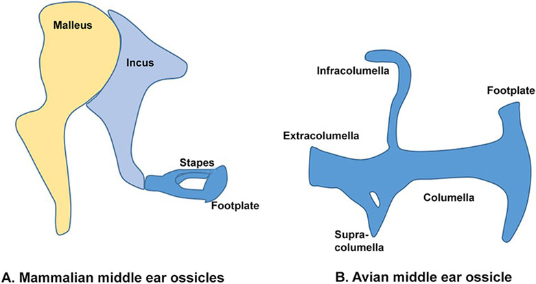
The mammalian middle ear comprises a chain of ossicles, the malleus, incus, and stapes that act as an impedance matching device during the transmission of sound from the tympanic membrane to the inner ear. These ossicles are derived from cranial neural crest cells that undergo endochondral ossification and subsequently differentiate into their final functional forms. Defects that occur during middle ear development can result in conductive hearing loss. In this review, we summarize studies describing the crucial roles played by signaling molecules such as sonic hedgehog, bone morphogenetic proteins, fibroblast growth factors, notch ligands, and chemokines during the differentiation of neural crest into the middle ear ossicles. In addition to these cell-extrinsic signals, we also discuss studies on the function of transcription factor genes such as Foxi3, Tbx1, Bapx1, Pou3f4, and Gsc in regulating the development and morphology of the middle ear ossicles.

Keywords: columella; growth factors; incus; malleus; middle ear; neural crest cells; ossicle; stapes; transcription factors.


Figures

Figure 1:
Figure 1:. Comparison of the mammalian middle ear ossicles and avian columella.
(A) The mammalian middle ear ossicles consists of the malleus, incus and stapes. The stapes is connected to the oval window of the inner ear through the stapedial footplate. (B) The avian columella is homologous to the mammalian stapes. It consists of the extracolumella, infracolumella, supracolumella, columella and footplate.
Figure 2:
Figure 2:. Endoderm acts as a signaling center for developing neural crest derived middle ear ossicles.
Loss of endodermal Shh (Foxg1Cre;Shhlox/lox) function, or inactivation of SHH signaling by deleting Smo in neural crest cells (Wnt1-Cre;Smolox/lox) lead to loss of the malleus and incus condensation, but not the stapes (C, D). Similarly, loss of endodermal Bmp4 (Foxg1Cre;Bmp4lox/Tm1) function, or inactivation of TGF-β/BMP signaling in neural crest cells by deleting Smad4 in neural crest cells resulted in the loss of the stapes condensation, but not the malleus or incus condensations (E,F). M, malleus; I, incus; S, stapes; DA, Dorsal aorta; PE, pharyngeal endoderm. This figure is adopted from Ankamreddy et al., Development (2019) with permission.
Figure 3:
Figure 3:. Summary of middle ear defects observed in different lines of mutant mice.
The middle ear ossicle defects observed in various mouse mutants are summarized in comparison to the wild type ossicles (A). Deletion of genes result in the loss of all three middle ear ossicles including their associated parts such as the tympanic ring and gonial bone (B, I, M), or a subset of the ossicles such as loss of the malleus and incus (C), incus (K) or stapes (E, J). Deletion of some genes can result in malformation of middle ear ossicles, such as a malformed malleus (J, L, M, N), malformed incus (G), malformed stapes (D, F, G, H) or malformed tympanic ring and gonial bone (J, N). Inactivation of Hoxa2 resulted in a mirror image duplication of the malleus, incus, tympanic ring and gonial bone in place of the stapes and other second pharyngeal arch derivatives (E). These phenotypes are summarized in Table 1. Solid red and blue colors indicate normal bone and cartilages, respectively. Dotted red and blue colors indicate the absence of bone and cartilages, respectively.

References

    1. Basch ML, Brown RM 2nd, Jen HI, Groves AK. Where hearing starts: the development of the mammalian cochlea. J Anat. 2015. - PMC - PubMed
    1. Mallo M. Formation of the middle ear: recent progress on the developmental and molecular mechanisms. Dev Biol. 2001;231(2):410–419. - PubMed
    1. Amin S, Matalova E, Simpson C, Yoshida H, Tucker AS. Incudomalleal joint formation: the roles of apoptosis, migration and downregulation. BMC developmental biology. 2007;7:134. - PMC - PubMed
    1. Park HY, Han DH, Lee JB, Han NS, Choung YH, Park K. Congenital stapes anomalies with normal eardrum. Clinical and experimental otorhinolaryngology. 2009;2(1):33–38. - PMC - PubMed
    1. Vincent R, Wegner I, Derks LS, Grolman W. Congenital ossicular chain malformations with mobile stapes in children: Results in 17 cases. Laryngoscope. 2016;126(3):682–688. - PubMed

Publication types

MeSH terms