Review
. 2020 Dec;29(12):2348-2362.
doi: 10.1002/pro.3973. Epub 2020 Oct 24.

How bilayer properties influence membrane protein folding

Affiliations
Review

How bilayer properties influence membrane protein folding

Karolina Corin et al. Protein Sci. 2020 Dec.

Abstract

The question of how proteins manage to organize into a unique three-dimensional structure has been a major field of study since the first protein structures were determined. For membrane proteins, the question is made more complex because, unlike water-soluble proteins, the solvent is not homogenous or even unique. Each cell and organelle has a distinct lipid composition that can change in response to environmental stimuli. Thus, the study of membrane protein folding requires not only understanding how the unfolded chain navigates its way to the folded state, but also how changes in bilayer properties can affect that search. Here we review what we know so far about the impact of lipid composition on bilayer physical properties and how those properties can affect folding. A better understanding of the lipid bilayer and its effects on membrane protein folding is not only important for a theoretical understanding of the folding process, but can also have a practical impact on our ability to work with and design membrane proteins.

Keywords: lipids; membrane insertion; packing pressure; phospholipids; reconstitution; stability; topology.


Conflict of interest statement

K.C. declares no conflict of interest. J.U.B. has founded a company involved in the production of natural chemicals that bears no direct relation to this work.

Figures

FIGURE 1
FIGURE 1
The structures, properties, and bilayer‐forming tendencies of common membrane lipids. Lipids can have neutral (green), zwitterionic (blue), anionic (orange), or cationic (grey) head groups, and acyl chains of varying length (denoted R1 and R2). The glycerol backbone is colored red. Reprinted from Biochim Biophys Acta 1818(4), Dowhan W and Bogdanov M, Molecular genetic and biochemical approaches for defining lipid‐dependent membrane protein folding, 1,097–1,107, Copyright (2012), with permission from Elsevier 113
FIGURE 2
FIGURE 2
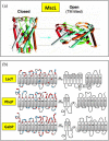
Examples of dramatic structural changes upon alterations in lipid composition. (a) Structural changes in MscL in response to changes in lipid composition. When MscL is reconstituted into 18:1 dioleoylphosphatidylcholine, the closed state (left) is observed. When lysophosphatidylcholine is added to one side of the bilayer the open conformation (right) is stabilized. 16 , 17 Adapted from Curr Opin Struct Biol 13(4), Perozo E and Rees DC, Structure and mechanism in prokaryotic mechanosensitive channels, 432–42, Copyright (2003), with permission from Elsevier. 17 (b) Topological flipping of LacY, PheP, and GabP in the presence and absence of PE. 47 , 48 , 54 , 57 The approximate locations of positively charged residues are shown with red dots, and the locations of negatively charged residues are shown with blue dots. Figure adapted from Refs. 48, 53, 54, 55, 56, 57, 113
FIGURE 3
FIGURE 3
Phase‐forming tendencies of lipids. Lipids with a cylindrical shape tend to form planar bilayer structures. Examples include phosphatidylcholine and phosphatidylserine. Lipids with an inverted cone shape tend to curve away from water and form micelles. Examples include lysophosphatidylcholine and other lysophospholipids. Lipids with a conical shape tend to curve toward water and form the inverted hexagonal phase. Examples include phosphatidylethanolamine and phosphatidic acid. Reprinted from FEBS Lett 593(17), Zhukovsky MA, Filograna A, Luini A, Corda D, Valente C, Phophatidic acid in membrane arrangements, 2,428–2,451, Copyright (2019), with permission from Wiley 115
FIGURE 4
FIGURE 4
A typical lateral pressure profile through a bilayer. Adding nonbilayer lipids to bilayer lipids increases the pressure in the chain region while decreasing the pressure in the head group region. Adding lipids with longer acyl chains can also increase the lateral chain pressure, while lysophospholipids can decrease the pressure. Figure adapted from Ref. 34
FIGURE 5
FIGURE 5
How bilayer pressure can shape membrane proteins or cause the bilayer to adapt to them. (a) Nonbilayer lipids can make an hourglass shape more stable. (b) The drive to shield hydrophobic side chains can either alter the protein conformation, the bilayer conformation, or both. Lipid shapes can facilitate or hinder bilayer adjustments
FIGURE 6
FIGURE 6
Lipid mixtures are necessary to simultaneously ensure proper folding, stability, insertion, and activity. LacY reconstitution, refolding, topology, and activity were assayed in different compositions of DOPE, DOPG, and DOPC, and the optimal concentrations for each individual parameter, as well as for all of them together, are shown. (a) Legend showing the relative fractions of DOPE, DOPG, and DOPC on each gridline. The corners labeled DOPE, DOPG, and DOPC represent samples with 100% of each lipid, respectively. The edges represent bilayer mixtures. For example, the edge between DOPE and DOPC represents samples made of those two lipids in varying percentages, where the orange numbers along the edge state the DOPE fraction. Each point or corner in the center of the large triangle represents samples with a mixture of all three lipids, with the numbers along the edge describing the exact composition at each point. (b) The reconstitution of LacY is optimal at high DOPG fractions, and impeded at low DOPE fractions. (c) The refolding of LacY is inhibited at high DOPE and DOPC fractions. (d) High DOPG fractions impede correct topological organization of LacY. (e) LacY transport activity is optimal at high DOPE fractions. (f) The lipid fractions at which LacY reconstitution, refolding, topology, and activity are all favorable, even if they are not all optimal. Only a subset of lipid compositions can support both proper protein folding and function. In (b)‐(e), each colored dot represents the results of an experiment at the indicated lipid composition. The color of the dot shows how well each lipid composition supports proper LacY structure and activity, with green dots representing optimal function or folding and red dots illustrating when the protein is most compromised. Adapted from Sci Rep 7(1):13056, Findlay HE and Booth PJ, The folding, stability, and function of lactose permease differ in their dependence on bilayer lipid composition, Copyright (2017) 64

References

    1. Casares D, Escribá PV, Rosselló CA. Membrane lipid composition: Effect on membrane and organelle structure, function and compartmentalization and therapeutic avenues. Int J Mol Sci. 2019;20:2167. - PMC - PubMed
    1. Sohlenkamp C, Geiger O. Bacterial membrane lipids: Diversity in structures and pathways. FEMS Microbiol Rev. 2016;40:133–159. - PubMed
    1. Dowhan W, Vitrac H, Bogdanov M. Lipid‐assisted membrane protein folding and topogenesis. Protein J. 2019;38:274–288. - PMC - PubMed
    1. Harayama T, Riezman H. Understanding the diversity of membrane lipid composition. Nat Rev Mol Cell Biol. 2018;19:281–296. - PubMed
    1. Shevchenko A, Simons K. Lipidomics: Coming to grips with lipid diversity. Nat Rev Mol Cell Biol. 2010;11:593–598. - PubMed

Publication types

MeSH terms