Review
. 2020 Oct 14;108(1):93-110.
doi: 10.1016/j.neuron.2020.09.003.

Ultrasound Technologies for Imaging and Modulating Neural Activity

Affiliations
Review

Ultrasound Technologies for Imaging and Modulating Neural Activity

Claire Rabut et al. Neuron. .

Abstract

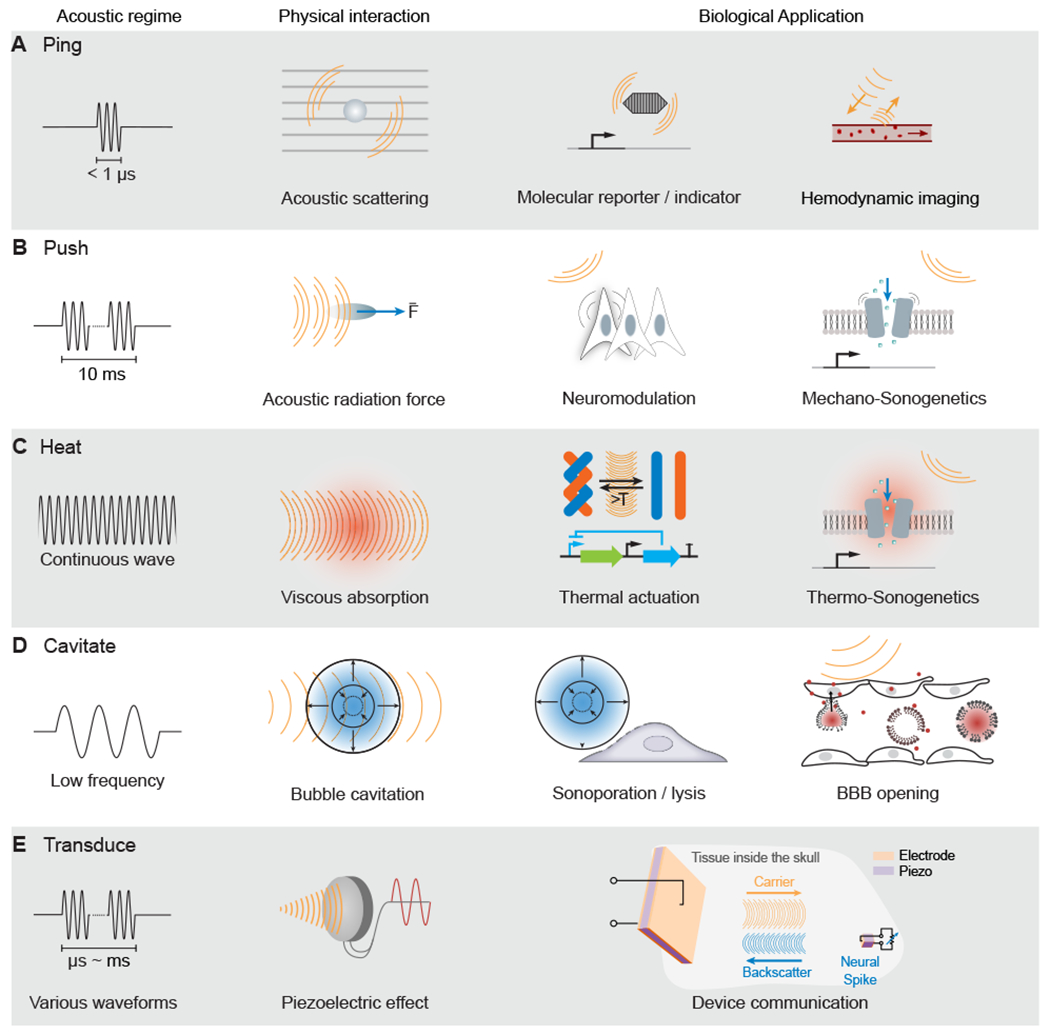
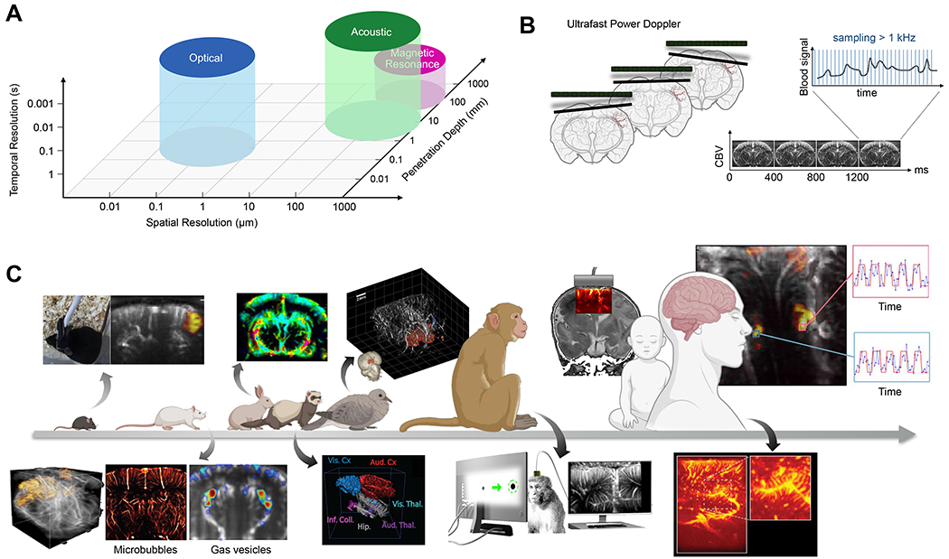
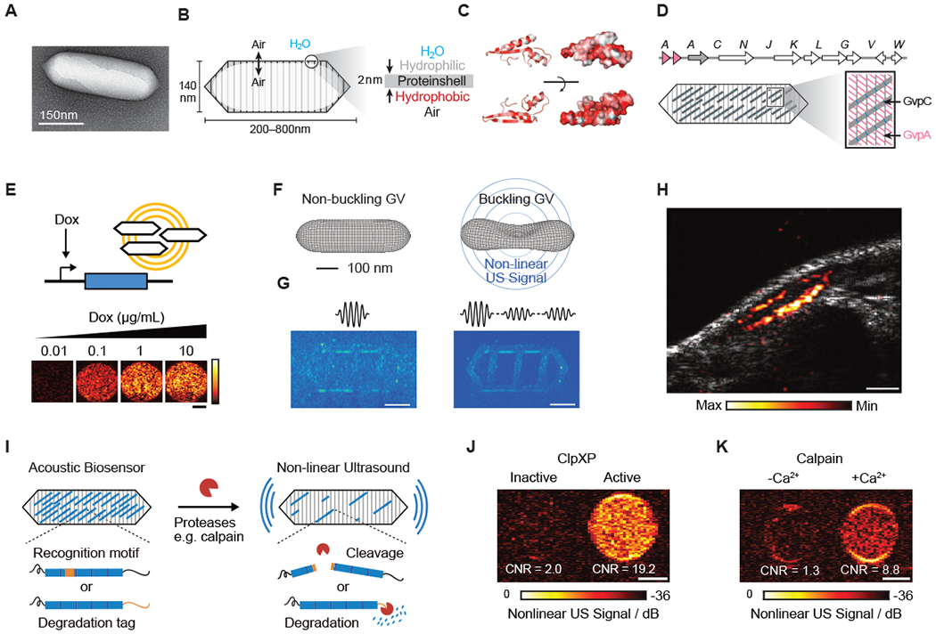
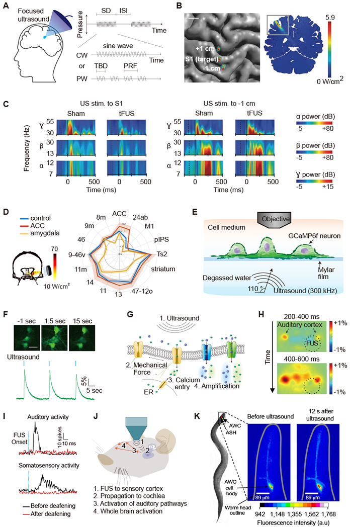
Visualizing and perturbing neural activity on a brain-wide scale in model animals and humans is a major goal of neuroscience technology development. Established electrical and optical techniques typically break down at this scale due to inherent physical limitations. In contrast, ultrasound readily permeates the brain, and in some cases the skull, and interacts with tissue with a fundamental resolution on the order of 100 μm and 1 ms. This basic ability has motivated major efforts to harness ultrasound as a modality for large-scale brain imaging and modulation. These efforts have resulted in already-useful neuroscience tools, including high-resolution hemodynamic functional imaging, focused ultrasound neuromodulation, and local drug delivery. Furthermore, recent breakthroughs promise to connect ultrasound to neurons at the genetic level for biomolecular imaging and sonogenetic control. In this article, we review the state of the art and ongoing developments in ultrasonic neurotechnology, building from fundamental principles to current utility, open questions, and future potential.

Keywords: focused ultrasound; functional imaging; molecular imaging; neuromodulation; neuroscience; sonogenetics; ultrasound.


Conflict of interest statement

Declaration of Interests The California Institute of Technology owns patents and patent applications related to some of the technologies described in this article.

Figures

Figure 1 -
Figure 1 -. Acoustic Regimes
(A) Ultrasound imaging involves the emission of brief pulses of sound and recording of backscattered echoes from materials such as molecular reporters and blood cells. (B) Sustained ultrasound application on the scale of milliseconds can generate mechanical forces, leading to cellular or molecular actuation. (C) Extended ultrasound can deposit thermal energy in tissues, which can activate temperature-dependent molecular function. (D) Ultrasound at lower frequencies can interact with bubbles to produce cavitation, leading to mechanical effects, such as blood-brain barrier (BBB) opening (A–D adapted from ​Maresca et al., 2018a​). (E) Ultrasound can communicate wirelessly with inorganic materials, such as millimeter-scale piezoelectric neural sensors (adapted from ​Seo et al., 2016​).
Figure 2 -
Figure 2 -. Functional Ultrasound Imaging.
(A) Approximate performance characteristics of common brain-imaging techniques. (B) The transmission of ultrasound plane wave in the brain allows a fast temporal sampling of the Doppler signal for highly sensitive measures of CBV variations (adapted from Deffieux et al., 2018). (C) Functional ultrasound imaging has been applied in many animal models and humans. Above the arrow: from left to right: functional imaging in awake mice (Tiran et al., 2017), dynamic changes in cerebral blood flow in rabbits undergoing cardiac arrest (Demené et al., 2018), 3D functional imaging of a pigeon (Rau et al., 2018), monitoring of cerebral activity through the ultrasound permeable anterior fontanel window in newborns (Demene et al., 2017), and intraoperative functional mapping to maximize tumor removal while preserving functional brain areas (Imbault et al., 2017) are shown. Below the arrow: from left to right: 3D functional imaging of rats using a matrix array probe (Rabut et at., 2019), ultrafast ultrasound localization microscopy allows sub-wavelength structural imaging of cerebral microvessels (Errico et al., 2015), enhancement of hemodynamic signal using gas vesicles (Maresca et al., 2020), 3D tonotopic mapping of the auditory pathway of awake ferrets (Bimbard et al.), detection of functional activation in awake monkeys during visual tasks (Dizeux et at, 2019), and rich vascular characteristics found in pre-resection tumors during intraoperative fUSI acquisition (Soloukey et al., 2020) are shown.
Figure 3 -
Figure 3 -. Biomolecular Ultrasound.
(A) Transmission electron microscopy (TEM) image of a gas vesicle (GV). (B) Diagram of a GV, showing the ability of gas to cross the shell, and the exclusion of water (left); physiochemical properties of the GV shell, namely the hydrophilic exterior that helps to solubilize the GV in the aqueous environment of the cell and the hydrophobic interior that prevents water from forming a liquid phase inside the GV (right). (C) Structural models of GvpA, the primary structural protein of the GV. Hydrophilic residues are colored white and hydrophobic residues red. The former can be seen to cluster on the convex side and the latter on the concave side. (D) (Top) Gene cluster from Anabaena flos-aquae encoding the formation of GVs, with each Gvp gene labeled. (Bottom) Diagram of a GV showing the relative contributions of the two main structural proteins to the structure of the GV (A-D from Maresca et al., 2018a). (E) Ultrasound images of human HEK293T cells in agarose gel expressing GVs under the control of a doxycycline (Dox)-inducible promoter. Scale bar, 1 mm (adapted from Farhadi et al., 2019). (F) Diagram showing the linear and nonlinear responses of GVs to ultrasound. (G) (Left) Ultrasound pulse sequence and corresponding image for a sample in the linear imaging mode. Tissue-mimicking linearly scattering particles and GVs are indistinguishable. (Right) The same sample imaged in nonlinear mode is shown, which detects nonlinearly scattering GVs, but not linearly scattering particles (F and G adapted with permission from Maresca et al., 2017) . (H) Ultrasound image of a GV-expressing tumor growing directly underneath the skin of a mouse. Scale bar, 1 mm (adapted from Farhadi et al., 2019). (I) Diagram of GV-based acoustic biosensor of protease activitiy (J) Representative ultrasound images of agarose phantoms containing acoustic biosensors sensing ClpXP activity. (K) Representative ultrasound images of agarose phantoms containing acoustic biosensor of calpain incubated with calpain and Ca2+ or calpain without Ca2+. (I-K adapted with permission from Lakshmanan et al., 2020). Scale bars for (J) and (K), 1 mm.
Figure 4 -
Figure 4 -. Ultrasonic Neuromodulation and Sonogenetics
(A) Illustration of focused ultrasound application to the brain and acoustic pulse parameters. CW, continuous wave; ISI, inter stimulus interval; PRF, pulse repetition frequency; PW, pulsed wave; SD, sonication duration; TBD, tone burst duration. (B) Top-down view of the brain showing acoustic intensity field of ultrasound beam targeting the S1 and sites 1 cm anterior (+1 cm) and posterior (−1 cm). (C) Time-frequency plots showing the power of evoked neural oscillations in the α, β, γ frequency bands in relation to the onset of ultrasound (dashed vertical line) and median nerve stimulation (solid vertical line) for sham and ultrasound treatment condition (B and C adapted with permission from Legon et al., 2014). (D) Left: acoustic intensity field targeting amygdala area of the primate brain. Right: functional connectivity fingerprint shows the strength of activity coupling between amygdala and other areas in control (blue), after ultrasound to amygdala (yellow), and after ultrasound to anterior cingulate cortex (ACC, red; adapted with permission from Folloni et al., 2019). (E) Diagram of experimental setup for in vitro neuronal ultrasound stimulation under acoustically realistic conditions. (F) GCaMP6f calcium signals in cultured neurons in response to ultrasound stimulation. Scale bar, 30 μm. (G) Biomolecular mechanisms of ultrasonic neuromodulation (E-G adapted with permission from Yoo et al., 2020). (H) Indirect excitation of mouse auditory cortex by FUS application to the visual cortex followed by widespread cortical response, as observed with wide-field calcium imaging (adapted with permission from Sato et al., 2018). (I) Neuronal responses to FUS recorded from guinea pig auditory or somatosensory cortex before and after deafening. (J) Mechanisms of indirect ultrasonic neuromodulation, where propagating ultrasound waves vibrate the cochlea, activating both auditory and non-auditory ascending pathways, leading to widespread activation. Deafening eliminates ultrasound-evoked multiunit sensory activity (I and J adapted from with permission from Guo et al., 2018). (K) Ultrasound activation of calcium signal in a C. elegans neuron engineered to express TRP-4 (adapted with permission from Ibsen et al., 2015).
Figure 5 -
Figure 5 -. Acoustically Targeted Pharmacology and Chemogenetics
(A) Illustration of microbubble-mediated blood brain barrier (BBB) opening by focused ultrasound. (B) Left: illustration of drug release from propofol-loaded nanoemulsions (adapted from Airan et al., 2017). Middle: illustration of ultrasound focal zone. Right: Fluorodeoxyglucose Positron emission tomography (FDG PET) images captured during sonication with or without propofol-loaded nanoparticle administration are shown. Scale bar, 5 mm (adapted from Wang et al., 2018). (C) Schematic of acoustically targeted chemogenetics (ATAC) paradigm. (D) Representative MRI scan indicating the site of the BBB opening (top, scale bar: 1 mm) and immunostaining imaging (bottom, scale bar: 200 μm). AAVs encoding hMsDq-mCherry were selectively delivered to the left SNc/VTA. The targeted neurons were excited by clozapine-N-oxide (CNO) ( C and D adapted with permission from Szablowski et al., 2018).

References

    1. Aaslid R, Markwalder T-M, and Nornes H (1982). Noninvasive transcranial Doppler ultrasound recording of flow velocity in basal cerebral arteries. J. Neurosurg 57, 769–774. - PubMed
    1. Airan RD, Meyer RA, Ellens NPK, Rhodes KR, Farahani K, Pomper MG, Kadam SD, and Green JJ (2017). Noninvasive Targeted Transcranial Neuromodulation via Focused Ultrasound Gated Drug Release from Nanoemulsions. Nano Lett. 17, 652–659. - PMC - PubMed
    1. Andreoni A, Davis CMO, and Tian L (2019). Measuring brain chemistry using genetically encoded fluorescent sensors. Curr. Opin. Biomed. Eng 12, 59–67.
    1. Aydin A-K, Haselden WD, Goulam Houssen Y, Pouzat C, Rungta RL, Demené C, Tanter M, Drew PJ, Charpak S, and Boido D (2020). Transfer functions linking neural calcium to single voxel functional ultrasound signal. Nat. Commun 11, 2954. - PMC - PubMed
    1. Bartelle BB, Barandov A, and Jasanoff A (2016). Molecular fMRI. J. Neurosci 36, 4139–4148. - PMC - PubMed

Publication types