. 2020 Nov 19;80(4):712-725.e5.
doi: 10.1016/j.molcel.2020.09.024. Epub 2020 Oct 14.

Cancer-Associated Gain-of-Function Mutations Activate a SWI/SNF-Family Regulatory Hub

Affiliations

Cancer-Associated Gain-of-Function Mutations Activate a SWI/SNF-Family Regulatory Hub

Cedric R Clapier et al. Mol Cell. .

Abstract

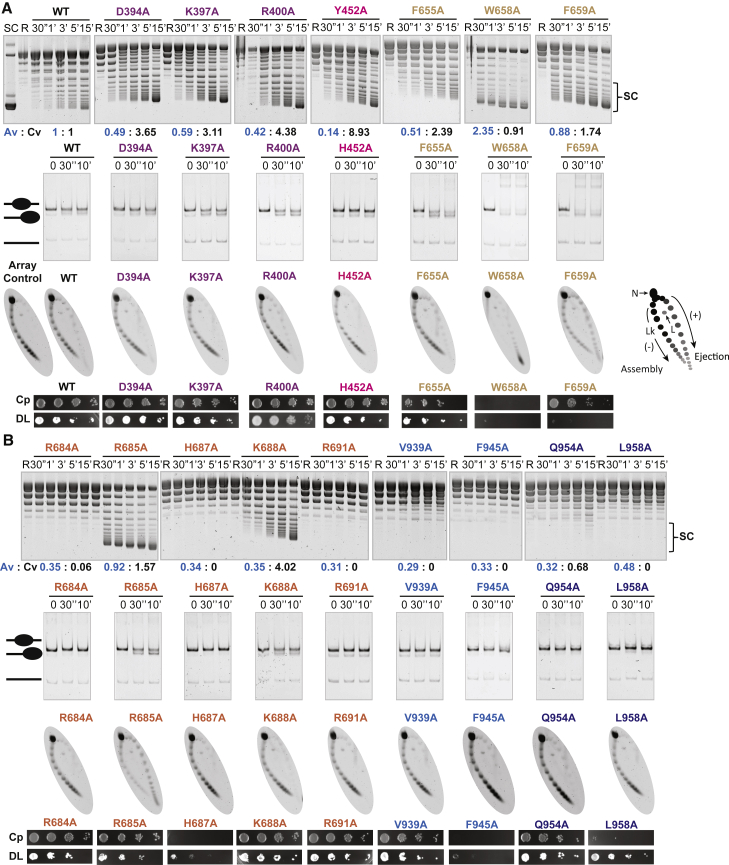
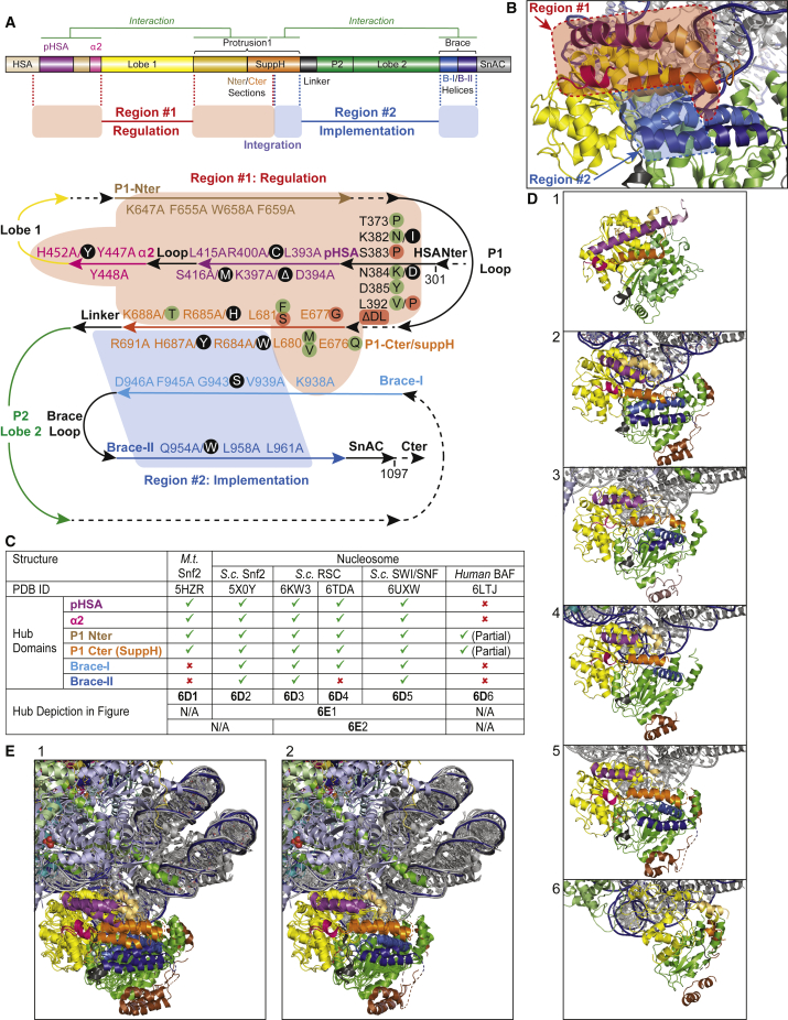
SWI/SNF-family remodelers (BAF/PBAF in mammals) are essential chromatin regulators, and mutations in human BAF/PBAF components are associated with ∼20% of cancers. Cancer-associated missense mutations in human BRG1 (encoding the catalytic ATPase) have been characterized previously as conferring loss-of-function. Here, we show that cancer-associated missense mutations in BRG1, when placed into the orthologous Sth1 ATPase of the yeast RSC remodeler, separate into two categories: loss-of-function enzymes, or instead, gain-of-function enzymes that greatly improve DNA translocation efficiency and nucleosome remodeling in vitro. Our work identifies a structural "hub," formed by the association of several Sth1 domains, that regulates ATPase activity and DNA translocation efficiency. Remarkably, all gain-of-function cancer-associated mutations and all loss-of-function mutations physically localize to distinct adjacent regions in the hub, which specifically regulate and implement DNA translocation, respectively. In vivo, only gain-of-function cancer-associated mutations conferred precocious chromatin accessibility. Taken together, we provide a structure-function mechanistic basis for cancer-associated hyperactivity.

Keywords: BAF; BRG1; DNA accessibility; RSC; STH1; SWI/SNF; cancer; chromatin remodeling; nucleosome.


Conflict of interest statement

Declaration of Interests The authors declare no competing interests.

Figures

None
Graphical abstract
Figure 1
Figure 1
An Integrative Regulatory Hub within the ATPase/Translocase of SWI/SNF Remodelers (A) Top: structure of the Snf2 ATPase/DNA-translocase (residues 666–1,400) bound to nucleosome (PDB: 5X0Y) (Liu et al., 2017). As Sth1 is highly similar to Snf2 over this region, it serves as a structural model. Middle: magnification and rotation of the top panel, to reorient and focus on the structural hub, viewed from the perspective of the schematic eye. The red arrow (top panel) and red dot (below the top panel) depict the axis of rotation, and the black arrow the direction of rotation. The helices of the integrative regulatory hub (purple, post-HSA; fuchsia, α2; beige, protrusion 1 N-terminal section; orange, protrusion 1 C-terminal section (SuppH); light blue, brace-I; dark blue, brace-II) flank the ATPase/DNA-translocase lobes (yellow, lobe 1; green, protrusion 2 and lobe 2). Bottom: linear schematic of the Snf2/Sth1 ATPase/DNA translocase subunit, depicting the conserved regulatory regions and their interactions (green connectors). This schematic includes the HSA domain, which forms a long helix that binds ARPs (Schubert et al., 2013), but the HSA is not present in the Snf2 structure, above. (B) Schematic representation of the hub, depicting all the residues investigated in this study, with each domain color-coded. Arrows depict domain direction (Nter-to-Cter), whereas dashed lines depict physical connectors/linkers between domains. (C) Table listing 11 human BRG1 residues mutated in cancer, with the number of samples and mutations, tissue distribution, and histology subtype (COSMIC database) (Tate et al., 2019) and their corresponding residue in Sth1. Domains to which these mutations map depicted on the right.
Figure 2
Figure 2
Alanine Substitutions Either Enhance or Abolish DNA Translocation, Coupling, and Nucleosome Sliding, Revealing Two Distinct Functional Regions within the Hub (A) Comparative impact of alanine substitutions within region 1. First row: comparative Tet-tethered DNA translocation activity measured by the accumulation of plasmid supercoiled (SC) topoisomers. Sth1 is tethered to the TetO containing plasmid via fusion to the DNA-binding domain of TetR (forming TetR-Sth1301–1097). Translocation along the DNA backbone creates positive supercoils in front of the translocase, and negative supercoils behind. E. coli Topoisomerase I (present in the reaction) relaxes only negative supercoils; thus, translocation yields one positive supercoil/10 bp translocation. The plasmid-stimulated ATPase activity/value (blue text, Av) and coupling value (black text, Cv, expressed as supercoiling/ATP hydrolyzed) and normalized to WT Sth1 (thus, Av and Cv values for WT Sth1 are 1.0). SC, highly supercoiled topoisomers; R, relaxed plasmid. Second row: comparative sliding of mononucleosomes (601 positioning DNA, 200 bp). Third row: comparative nucleosome ejection in a closed circular array format. At right: schematic of the principle of the nucleosome array ejection assay, with SC plasmid (topoisomer) distribution revealed by a 2D gel. Lk, linking number; N, nicked; L, linear. In sliding and ejection assays, the enzyme:nucleosome molar ratio is 1:2, and representative gels from multiple experiments are shown. Fourth row: comparative genetic assessment (complementation [Cp] and dominant-lethality [DL]) conferred by expression of particular Sth1 derivatives. Ability to complement assessed by expressing transformed Sth1 derivatives (regulated by WT STH1 promoter) in cells lacking genomic WT STH1. DL status assessed by expression using the methionine-regulated MET25 promoter, in the presence of WT STH1. Each panel displays a dilution series of yeast cells on plate media, testing growth ability. One representative of three replicates is shown. Mutations are color-coded according to the domains they belong to in the hub, as in Figure 1. (B) Comparative impact of alanine substitutions in the SuppH helix and region 2. All assays performed and results depicted as in (A).
Figure 3
Figure 3
Mutations that Suppress arpΔ Mutations or Exhibit Dominant Lethality Map within Region 1 (A) Mutations that suppress arpΔ mutations (mra mutants) increase coupling and DNA translocation, complement the loss of WT Sth1, and map to region 1. First row: comparative ATPase and DNA translocation assays performed using Sth1 derivatives harboring the 10 arpΔ-suppressing mra mutations (green), five clustered in the HSA, and five clustered in the SuppH of protrusion 1 (Szerlong et al., 2008). Second row: comparative genetic assessment as in Figure 2A. (B) Mutations that confer dominant lethality increase ATPase activity and DNA translocation fail to complement the loss of WT Sth1 and map to region 1. First row: comparative ATPase and DNA translocation assays performed using the five DL Sth1 mutants (red) located in the HSA and the SuppH of protrusion 1. Second row: comparative genetic assessment as in Figure 2A.
Figure 4
Figure 4
Cancer-Associated Missense Mutations Either Enhance or Abolish DNA Translocation, Coupling, and Nucleosome Sliding, Confirming Two Distinct Functional Regions within the Hub (A) Cancer-associated missense mutations within regulatory region 1 enhance DNA translocation coupling and nucleosome sliding. All assays performed and results depicted as in Figure 2A. (B) Cancer-associated missense mutations in the α2 helix, SuppH helix, and region 2 reveal the interface between regulatory region 1 and the implementation region 2. All assays performed and results depicted as in Figure 2A.
Figure 5
Figure 5
Gain-of-Function Cancer-Associated Mutations and Dominant Lethal Mutations Open Chromatin Moderately and Extensively, Respectively (A) Principle component analysis of ATAC-seq data partitions Sth1 hub mutants into three clusters. A modified ATAC-seq protocol (employing crosslinking) was applied to a subset of hub-localized sth1 mutants from three categories: (1) loss-of-function cancer-associated mutants (Q954W, H687Y, and R684W) from region 2, (2) gain-of-function mra (N384K) or cancer-associated (R400C and R685H) mutants from region 1, and (3) dominant lethal mutants (L681F, L392P, and W658A) from region 1. All Sth1 derivatives were expressed using a MET25 inducible promoter. The mean log2 fold ratio of signal over background was collected in a 1-kb window (−500 to +500 bp) of protein-coding gene TSS, and the first two principal components were plotted for the top 1,000 variant genes. Phenotypic categories are circled for emphasis. (B) Genome-wide impact of Sth1 hub mutations on chromatin structure at promoters. Top: a hierarchical clustering tree/dendrogram derived from the mean accessibility heatmap over 1 kb of all TSS (Figure S4C) shows that the sth1 mutants cluster into three categories. Middle: heatmaps of log2 fold ratio of DNA accessibility signal over genomic background at all promoter TSS (1 kb window, −500 to +500 bp) arranged in descending order of promoter NDR length. The sth1 mutants within each category appear similar, while the three categories display a progressive impact. Bottom: log2 mean profile of DNA accessibility ratio over 20 bp bins at all TSS (1-kb window, −500 to +500 bp) plotted to depict changes in accessibility between mutants from the three categories. (C) Genome browser snapshots depicting the impact of Sth1 mutants on chromatin openness. Accessibility coverage (log2 RPM coverage) across two representative chromosomal regions is shown. All tracks are log scaled to the same y axis.
Figure 6
Figure 6
The Domains Flanking the ATPase/DNA Translocase Lobes Form a Conserved Integrative Hub with a Regulatory Region 1 Where Cancer-Associated Gain-of-Function Mutations Map (A) Top: linear schematic depicting the functional roles and physical interaction of Sth1 hub domains. The regions flanking the two RecA-like lobes associate structurally to form an integrative regulatory hub comprised of two distinct regions: region 1 is formed by the HSA, the post-HSA, and the Nter and parts of the Cter helices (including one side of SuppH) of protrusion 1, and is involved in the regulation of coupling (light red connected boxes); and region 2, formed by one side of SuppH helix of protrusion 1 and the braces, involved in the implementation of coupling (light blue connected boxes). Remarkably, the Cter helix of protrusion 1 (termed SuppH) appears to be split between region 1 and region 2, integrating the regulation and directing its proper implementation. Bottom: detailed schematic model of the Sth1 integrative regulatory hub. All domains and mutations investigated are depicted, revealing two functional regions: region 1 (light red shape) involved in DNA translocation/coupling regulation and harboring gain-of-function mutations (cancer-associated missense mutations [black dots] and alanine substitutions), the mra mutations (green dots), and the DL mutations (red dots), and region 2 (light blue shape) involved in the implementation of coupling and harboring loss-of-function mutations (cancer-associated missense mutations [(black dots] and particular alanine substitutions). (B) Magnification of the integrative regulatory hub of Snf2 depicted as in Figure 1A, with the two revealed functional regions highlighted: region 1 (light red shape) and region 2 (light blue shape). (C) Comparison of structural hub domains visible (green tick symbol) or not built in the structural model (red cross symbol) in the structures of various remodelers (structure names and PDB IDs mentioned) and their depiction in (D). (D) Gallery of individual structural hubs from various remodelers aligned. (1) Myceliophthora thermophila Snf2 (PDB: 5HZR) (Xia et al., 2016). (2) S. cerevisiae Snf2 ATPase bound to a nucleosome (PDB: 5X0Y) (Liu et al., 2017). (3 and 4) S. cerevisiae RSC bound to a nucleosome (PDB: 6KW3 and 6TDA, respectively) (Wagner et al., 2020; Ye et al., 2019). (5): S. cerevisiae SWI/SNF bound to a nucleosome (PDB: 6UXW) (Han et al., 2020). (6) Human BAF bound to a nucleosome (PDB: 6LTJ) (He et al., 2020). (E) Structural alignment of various remodeler-nucleosome complexes (oriented as in D) based on the alignment of their respective histone octamers highlighting the structural conservation of the different domains forming the hub. (1) Alignment of S. cerevisiae Snf2 ATPase (PDB: 5X0Y), RSC complex (PDB: 6KW3 and 6TDA), and SWI/SNF complex (PDB: 6UXW), each bound to a nucleosome. (2) As in (1) but without the Snf2 ATPase structure.

References

    1. Baker R.W., Reimer J.M., Carman P.J., Arakawa T., Dominguez R., Leschziner A.E. Structural insights into assembly and function of the RSC chromatin remodeling complex. bioRxiv. 2020 doi: 10.1101/2020.1103.1124.006361. - DOI - PMC - PubMed
    1. Bartholomew B. Regulating the chromatin landscape: structural and mechanistic perspectives. Annu. Rev. Biochem. 2014;83:671–696. - PMC - PubMed
    1. Becker P.B., Workman J.L. Nucleosome remodeling and epigenetics. Cold Spring Harb. Perspect. Biol. 2013;5:a017905. - PMC - PubMed
    1. Buenrostro J.D., Wu B., Chang H.Y., Greenleaf W.J. ATAC-seq: A Method for Assaying Chromatin Accessibility Genome-Wide. Curr. Protoc. Mol. Biol. 2015;109:21–29. - PMC - PubMed
    1. Bultman S.J., Gebuhr T.C., Magnuson T. A Brg1 mutation that uncouples ATPase activity from chromatin remodeling reveals an essential role for SWI/SNF-related complexes in beta-globin expression and erythroid development. Genes Dev. 2005;19:2849–2861. - PMC - PubMed

Publication types

MeSH terms