

Epithelial-mesenchymal transition of absorptive enterocytes and depletion of Peyer's patch M cells after PEDV infection
- PMID: 33059319
- PMCID: PMC7548064
- DOI: 10.1016/j.virol.2020.08.018
Epithelial-mesenchymal transition of absorptive enterocytes and depletion of Peyer's patch M cells after PEDV infection
Abstract
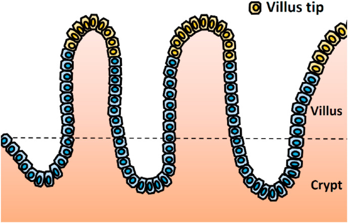
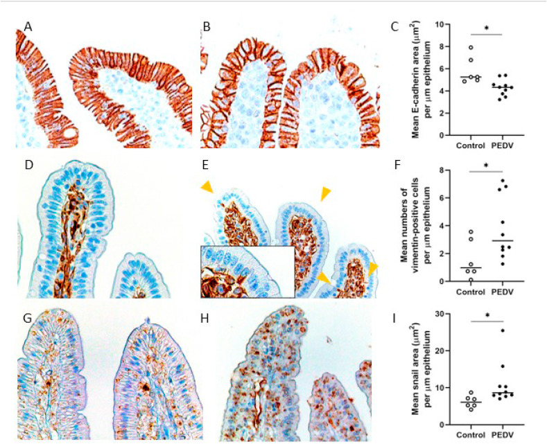
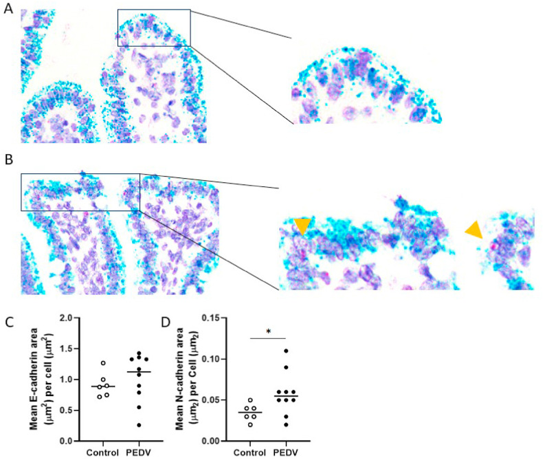
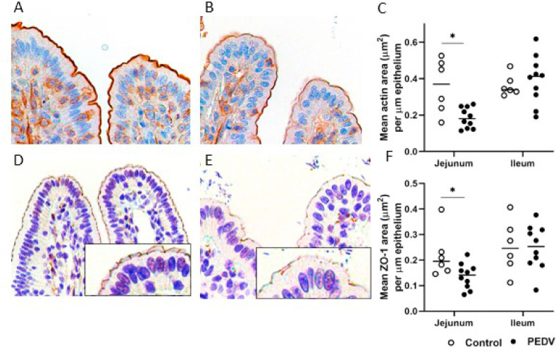
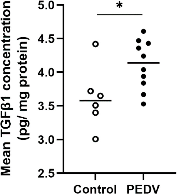
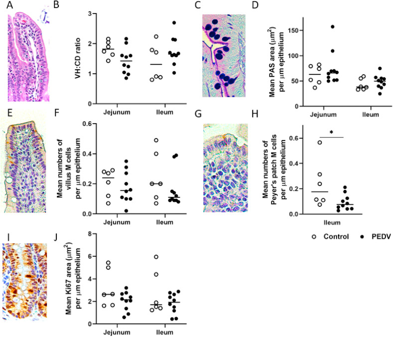
This study focused on intestinal restitution including phenotype switching of absorptive enterocytes and the abundance of different enterocyte subtypes in weaned pigs after porcine epidemic diarrhea virus (PEDV) infection. At 10 days post-PEDV-inoculation, the ratio of villus height to crypt depth in both jejunum and ileum had restored, and the PEDV antigen was not detectable. However, enterocytes at the villus tips revealed epithelial-mesenchymal transition (EMT) in the jejunum in which E-cadherin expression decreased while expression of N-cadherin, vimentin, and Snail increased. Additionally, there was reduced expression of actin in microvilli and Zonula occludens-1 (ZO-1) in tight junctions. Moreover, the protein concentration of transforming growth factor β1 (TGFβ1), which mediates EMT and cytoskeleton alteration, was increased. We also found a decreased number of Peyer's patch M cells in the ileum. These results reveal incomplete restitution of enterocytes in the jejunum and potentially impaired immune surveillance in the ileum after PEDV infection.
Keywords: Enteric coronavirus; Epithelial-mesenchymal transition; Intestine; Microfold cell; Weaned pig.
Copyright © 2020 Elsevier Inc. All rights reserved.
Conflict of interest statement
The author(s) declare no potential conflicts of interest with respect to the research, authorship, and/or publication of this article.
Figures
Similar articles
-
Alterations in Intestinal Innate Mucosal Immunity of Weaned Pigs During Porcine Epidemic Diarrhea Virus Infection.Vet Pathol. 2020 Sep;57(5):642-652. doi: 10.1177/0300985820932140. Epub 2020 Jun 17. Vet Pathol. 2020. PMID: 32880235
-
Structural alteration of tight and adherens junctions in villous and crypt epithelium of the small and large intestine of conventional nursing piglets infected with porcine epidemic diarrhea virus.Vet Microbiol. 2015 Jun 12;177(3-4):373-8. doi: 10.1016/j.vetmic.2015.03.022. Epub 2015 Mar 30. Vet Microbiol. 2015. PMID: 25843943 Free PMC article.
-
Comparative pathogenesis of US porcine epidemic diarrhea virus (PEDV) strain PC21A in conventional 9-day-old nursing piglets vs. 26-day-old weaned pigs.Vet Microbiol. 2015 Jul 9;178(1-2):31-40. doi: 10.1016/j.vetmic.2015.04.022. Epub 2015 Apr 30. Vet Microbiol. 2015. PMID: 25939885 Free PMC article.
-
Epidemiology of porcine epidemic diarrhea virus among Chinese pig populations: A meta-analysis.Microb Pathog. 2019 Apr;129:43-49. doi: 10.1016/j.micpath.2019.01.017. Epub 2019 Jan 23. Microb Pathog. 2019. PMID: 30682525
-
Three Main Inducers of Alphacoronavirus Infection of Enterocytes: Sialic Acid, Proteases, and Low pH.Intervirology. 2018;61(2):53-63. doi: 10.1159/000492424. Epub 2018 Sep 3. Intervirology. 2018. PMID: 30176660 Free PMC article. Review.
Cited by
-
The role of innate immune responses against two strains of PEDV (S INDEL and non-S INDEL) in newborn and weaned piglets inoculated by combined orogastric and intranasal routes.Front Immunol. 2025 Jun 16;16:1584785. doi: 10.3389/fimmu.2025.1584785. eCollection 2025. Front Immunol. 2025. PMID: 40589734 Free PMC article.
-
LncRNA446 Regulates Tight Junctions by Inhibiting the Ubiquitinated Degradation of Alix after Porcine Epidemic Diarrhea Virus Infection.J Virol. 2023 Mar 30;97(3):e0188422. doi: 10.1128/jvi.01884-22. Epub 2023 Feb 15. J Virol. 2023. PMID: 36790206 Free PMC article.
-
Lectin histochemistry in the small intestines of piglets naturally infected with porcine epidemic diarrhea virus.J Vet Sci. 2024 Sep;25(5):e66. doi: 10.4142/jvs.24179. J Vet Sci. 2024. PMID: 39363654 Free PMC article.
-
The Oral Inactivated Porcine Epidemic Diarrhea Virus Presenting in the Intestine Induces Mucosal Immunity in Mice with Alginate-Chitosan Microcapsules.Animals (Basel). 2023 Feb 28;13(5):889. doi: 10.3390/ani13050889. Animals (Basel). 2023. PMID: 36899746 Free PMC article.
-
Yersinia pseudotuberculosis YopE prevents uptake by M cells and instigates M cell extrusion in human ileal enteroid-derived monolayers.Gut Microbes. 2021 Jan-Dec;13(1):1988390. doi: 10.1080/19490976.2021.1988390. Gut Microbes. 2021. PMID: 34793276 Free PMC article.
References
-
- Albers T.M., Lomakina I., Moore R.P. Structural and functional roles of cytoskeletal proteins during repair of native Guinea pig intestinal epithelium. Cell Biol. Int. 1996;20:821–830. - PubMed
-
- Chen Q., Gauger P.C., Stafne M.R., Thomas J.T., Madson D.M., Huang H., Zheng Y., Li G., Zhang J. Pathogenesis comparison between the United States porcine epidemic diarrhoea virus prototype and S-INDEL-variant strains in conventional neonatal piglets. J. Gen. Virol. 2016;97:1107–1121. - PubMed
-
- Chen X., Bode A.M., Dong Z., Cao Y. The epithelial-mesenchymal transition (EMT) is regulated by oncoviruses in cancer. Faseb. J. 2016;30:3001–3010. - PubMed
-
- Chen Y.-M., Helm E., Gabler N., Hostetter J., Burrough E. Alterations in intestinal innate mucosal immunity of weaned pigs during porcine epidemic diarrhea virus infection. Vet. Pathol. 2020;57:642–652. - PubMed
Publication types
MeSH terms
Substances
LinkOut - more resources
Full Text Sources
Other Literature Sources
Research Materials
