. 2020 Oct 15;17(1):305.
doi: 10.1186/s12974-020-01967-2.

Forsythoside B attenuates memory impairment and neuroinflammation via inhibition on NF-κB signaling in Alzheimer's disease

Affiliations

Forsythoside B attenuates memory impairment and neuroinflammation via inhibition on NF-κB signaling in Alzheimer's disease

Fan'ge Kong et al. J Neuroinflammation. .

Abstract

Background: Neuroinflammation is a principal element in Alzheimer's disease (AD) pathogenesis, so anti-inflammation may be a promising therapeutic strategy. Forsythoside B (FTS•B), a phenylethanoid glycoside isolated from Forsythiae fructus, has been reported to exert anti-inflammatory effects. However, no studies have reported whether the anti-inflammatory properties of FTS•B have a neuroprotective effect in AD. In the present study, these effects of FTS•B were investigated using amyloid precursor protein/presenilin 1 (APP/PS1) mice, BV-2 cells, and HT22 cells.

Methods: APP/PS1 mice were administered FTS•B intragastrically for 36 days. Behavioral tests were then carried out to examine cognitive functions, including the Morris water maze, Y maze, and open field experiment. Immunohistochemistry was used to analyze the deposition of amyloid-beta (Aβ), the phosphorylation of tau protein, and the levels of 4-hydroxynonenal, glial fibrillary acidic protein, and ionized calcium-binding adapter molecule 1 in the hippocampus. Proteins that showed marked changes in levels related to neuroinflammation were identified using proteomics and verified using enzyme-linked immunosorbent assay and western blot. BV-2 and HT22 cells were also used to confirm the anti-neuroinflammatory effects of FTS•B.

Results: In APP/PS1 mice, FTS•B counteracted cognitive decline, ameliorated the deposition of Aβ and the phosphorylation of tau protein, and attenuated the activation of microglia and astrocytes in the cortex and hippocampus. FTS•B affected vital signaling, particularly by decreasing the activation of JNK-interacting protein 3/C-Jun NH2-terminal kinase and suppressing WD-repeat and FYVE-domain-containing protein 1/toll-like receptor 3 (WDFY1/TLR3), further suppressing the activation of nuclear factor-κB (NF-κB) signaling. In BV-2 and HT22 cells, FTS•B prevented lipopolysaccharide-induced neuroinflammation and reduced the microglia-mediated neurotoxicity.

Conclusions: FTS•B effectively counteracted cognitive decline by regulating neuroinflammation via NF-κB signaling in APP/PS1 mice, providing preliminary experimental evidence that FTS•B is a promising therapeutic agent in AD treatment.

Keywords: Alzheimer’s disease; Forsythoside B; NF-κB; Neuroinflammation.


Conflict of interest statement

The authors declare that they have no competing interests.

Figures

Fig. 1
Fig. 1
FTS•B counteracted cognitive decline in APP/PS1 mice. (a) In the Morris water maze test, the path tracings were automatically recorded on the testing day. FTS•B reduced (b) the escape latency on the 4 testing days in APP/PS1 mice, as well as (c) the times of crossing the platform on the testing day. (d) The path tracings were automatically recorded in the Y maze test. (e) FTS•B failed to influence training latency, but (f) strongly reduced the testing latency in the Y maze test. (g) The path tracings in the open field test were automatically recorded. (h) FTS•B increased the distance in the surrounding area and (i) reduced the distance in the center area of APP/PS1 mice in the open field test. Data are expressed as the mean ± standard error (n = 8). #p < 0.05, ##p < 0.01, and ###p < 0.001 vs. WT mice. *p < 0.05 and **p < 0.01 vs. APP/PS1 mice. FTS•B, forsythoside B; APP/PS1, amyloid precursor protein/presenilin 1; WT, wild type
Fig. 2
Fig. 2
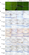
FTS•B ameliorated pathological alterations in the brains of APP/PS1 mice. FTS•B alleviated the deposition of Aβ in the brains of APP/PS1 mice, detected using (a) thioflavine S staining in the hippocampus and cortex (100×; scale bar: 100 μm; n = 3) and (b) immunohistochemistry in the hippocampus (40×; scale bar: 200 μm; 200×; scale bar: 50 μm; n = 3). FTS•B reduced the levels of (c) phospho-tau protein (n = 3), (d) 4-HNE (n = 3), and (e) GFAP (n = 3) in the hippocampus of APP/PS1 mice (40×; scale bar: 200 μm; 200×; scale bar: 50 μm). (f) FTS•B suppressed the enrichment of Iba1 around blood vessels in the hippocampus of APP/PS1 mice (n = 3; 40×; scale bar: 200 μm; 200×; scale bar: 50 μm). FTS•B, forsythoside B; APP/PS1, amyloid precursor protein/presenilin 1; 4-HNE, 4-hydroxynonenal; GFAP, glial fibrillary acidic protein; Iba1, ionized calcium-binding adapter molecule 1
Fig. 3
Fig. 3
Label-free proteomics performed to evaluate the protein changes caused by FTS•B administration in the hippocampus of APP/PS1 mice. (a) The heatmap shows differences in the cellular protein profiles in the hippocampus between WT and APP/PS1 mice with or without FTS•B administration. (b) The protein interaction image shows that significantly up- or downregulated proteins may be closely related to inflammation. FTS•B, forsythoside B; APP/PS1, amyloid precursor protein/presenilin 1; WT, wild type
Fig. 4
Fig. 4
The regulation of FTS•B on proteins related to neuroinflammation in the hippocampus of APP/PS1 mice. (a) FTS•B reduced the expression levels of JIP3 and the phosphorylation levels of JNK and APP. (b) FTS•B suppressed the expression levels of ELKS and the phosphorylation levels of IKK(α + β), IκBα, and NF-κB p65. (c) FTS•B enhanced the expression levels of WDFY1 and TLR3, as well as the phosphorylation levels of IRF3. (d) FTS•B reduced the expression levels of Iba1 and GFAP. Quantification data were normalized to GAPDH and corresponding total proteins (n = 3). They were reported as folds of the corresponding WT group. FTS•B, forsythoside B; APP/PS1, amyloid precursor protein/presenilin 1; JIP3, JNK-interacting protein 3; JNK, C-Jun NH2-terminal kinase; APP, amyloid-beta precursor protein; IKK, inhibitor of nuclear factor kappa-B kinase; IκBα, inhibitor of nuclear factor kappa-B alpha; NF-κB, nuclear factor-κB; WDFY1, WD-repeat and FYVE-domain-containing protein 1; TLR3, toll-like receptor 3; IRF3, interferon regulating factor 3; Iba1, ionized calcium-binding adapter molecule 1; GFAP, glial fibrillary acidic protein
Fig. 5
Fig. 5
FTS•B incubation regulated the levels of inflammatory cytokines in LPS-exposed BV-2 cells. (a) FTS•B alone or in co-incubation with LPS showed no effects on cell viability of BV-2 cells. Compared with LPS-alone exposed cells, FTS•B co-incubation strongly suppressed the release of (b) IL-6, (c) TNF-α, (d) iNOs, (e) NO, and (f) IL-1β in BV-2 cells, determined by ELISA. Data are expressed as mean ± standard error (n = 3). #p < 0.05 and ##p < 0.01 vs. non-treated cells. *p < 0.05, **p < 0.01, and ***p < 0.001 vs. LPS-alone exposed cells. FTS•B, forsythoside B; LPS, lipopolysaccharide; IL-6, interleukin 6; TNF-α, tumor necrosis factor α; iNOs, inducible nitric oxide synthase; NO, nitric oxide; IL-1β, interleukin 1β; ELISA, enzyme-linked immunosorbent assay
Fig. 6
Fig. 6
FTS•B reduced the microglia-mediated neurotoxicity induced by LPS. Mouse HT22 cells were treated using conditioned media from BV-2 microglia exposed to LPS for 24 h, with or without FTS•B. (a and b) The apoptosis rate and (c) cell viability were assessed. Data are expressed as mean ± standard error (n = 3). ###p < 0.001 vs. control group. *p < 0.05, **p < 0.01, and ***p < 0.001 vs. group treated with LPS. FTS•B, forsythoside B; LPS, lipopolysaccharide

References

    1. Caraci F, Santagati M, Caruso G, Cannavo D, Leggio GM, Salomone S, Drago F: New antipsychotic drugs for the treatment of agitation and psychosis in Alzheimer’s disease: focus on brexpiprazole and pimavanserin. F1000Research 2020, 9. - PMC - PubMed
    1. Bessey LJ, Walaszek A. Management of behavioral and psychological symptoms of dementia. Current Psychiatry Reports. 2019;21. - PubMed
    1. Huang L-K, Chao S-P, Hu C-J. Clinical trials of new drugs for Alzheimer disease. J Biomed Sci. 2020;27. - PMC - PubMed
    1. Toublet F-X, Lecoutey C, Lalut J, Hatat B, Davis A, Since M, Corvaisier S, Freret T, Santos JSO, Claeysen S, et al. Inhibiting acetylcholinesterase to activate pleiotropic prodrugs with therapeutic interest in Alzheimer’s disease. Molecules. 2019;24. - PMC - PubMed
    1. Medrano-Jimenez E, Jimenez-Ferrer Carrillo I, Pedraza-Escalona M, Ramirez-Serrano CE, Alvarez-Arellano L, Cortes-Mendoza J, Herrera-Ruiz M, Jimenez-Ferrer E, Zamilpa A, Tortoriello J, et al. Malva parviflora extract ameliorates the deleterious effects of a high fat diet on the cognitive deficit in a mouse model of Alzheimer’s disease by restoring microglial function via a PPAR-gamma-dependent mechanism. J Neuroinflammation. 2019;16. - PMC - PubMed

MeSH terms