

Molecular Imaging of Apoptosis in Atherosclerosis by Targeting Cell Membrane Phospholipid Asymmetry
- PMID: 33059832
- PMCID: PMC7654709
- DOI: 10.1016/j.jacc.2020.08.047
Molecular Imaging of Apoptosis in Atherosclerosis by Targeting Cell Membrane Phospholipid Asymmetry
Abstract
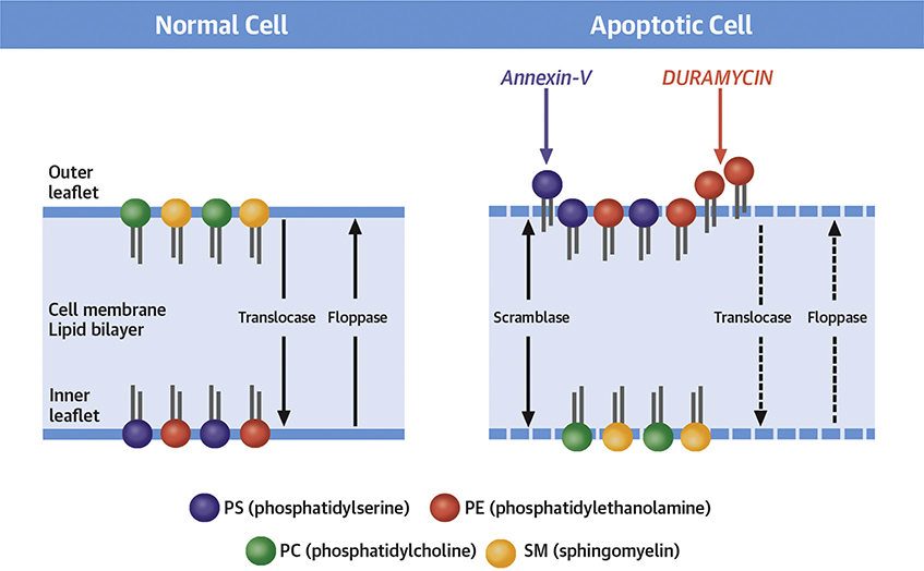
Background: Apoptosis in atherosclerotic lesions contributes to plaque vulnerability by lipid core enlargement and fibrous cap attenuation. Apoptosis is associated with exteriorization of phosphatidylserine (PS) and phosphatidylethanolamine (PE) on the cell membrane. Although PS-avid radiolabeled annexin-V has been employed for molecular imaging of high-risk plaques, PE-targeted imaging in atherosclerosis has not been studied.
Objectives: This study sought to evaluate the feasibility of molecular imaging with PE-avid radiolabeled duramycin in experimental atherosclerotic lesions in a rabbit model and compare duramycin targeting with radiolabeled annexin-V.
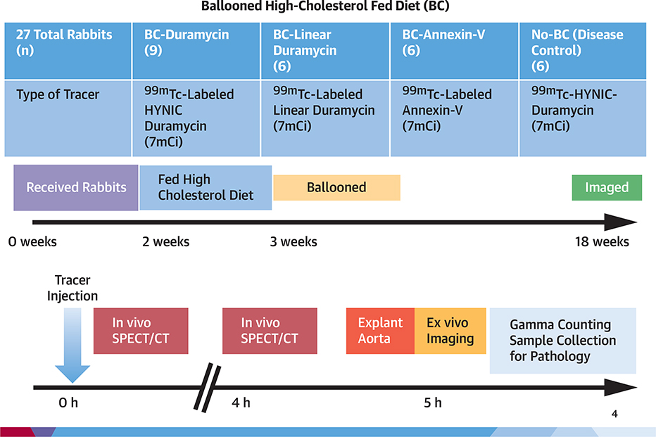
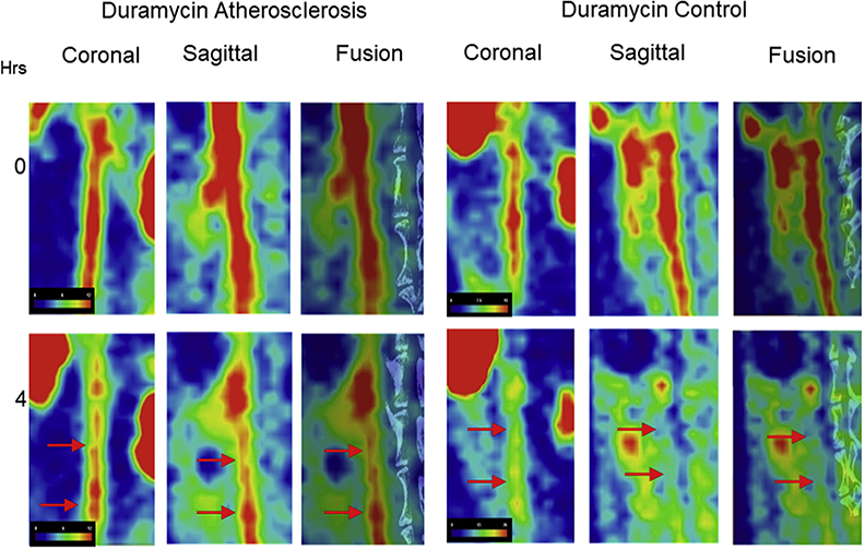
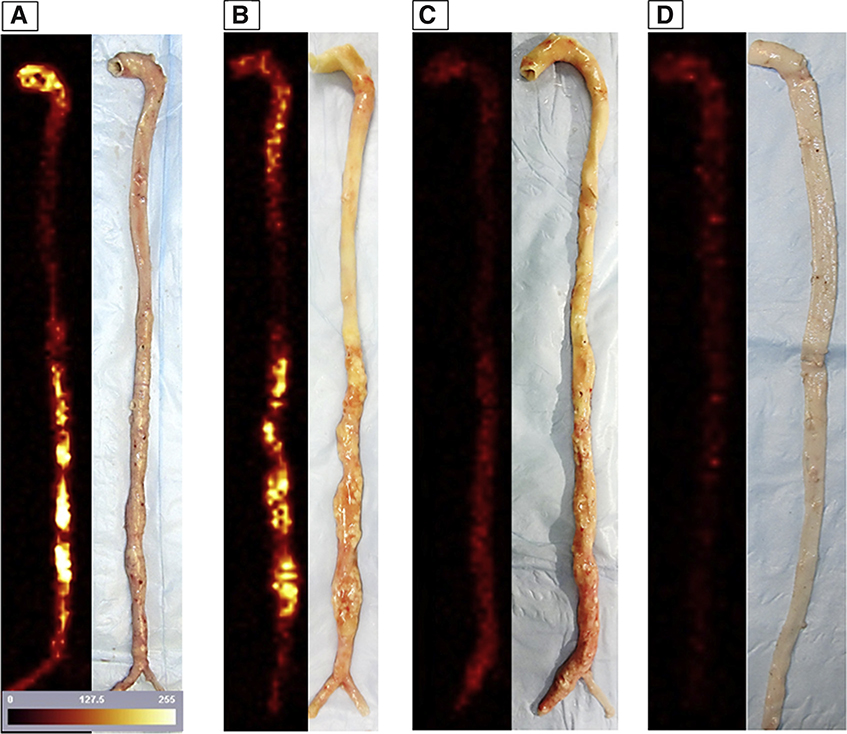
Methods: Of the 27 rabbits, 21 were fed high-cholesterol, high-fat diet for 16 weeks. Nine of the 21 rabbits received 99mTc-duramycin (test group), 6 received 99mTc-linear duramycin (duramycin without PE-binding capability, negative radiotracer control group), and 6 received 99mTc-annexin-V for radionuclide imaging. The remaining normal chow-fed 6 animals (disease control group) received 99mTc-duramycin. In vivo microSPECT/microCT imaging was performed, and the aortas were explanted for ex vivo imaging and for histological characterization of atherosclerosis.
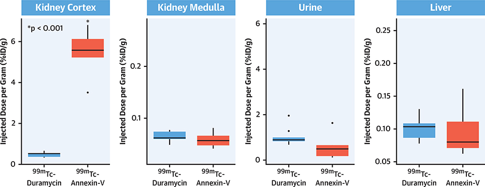
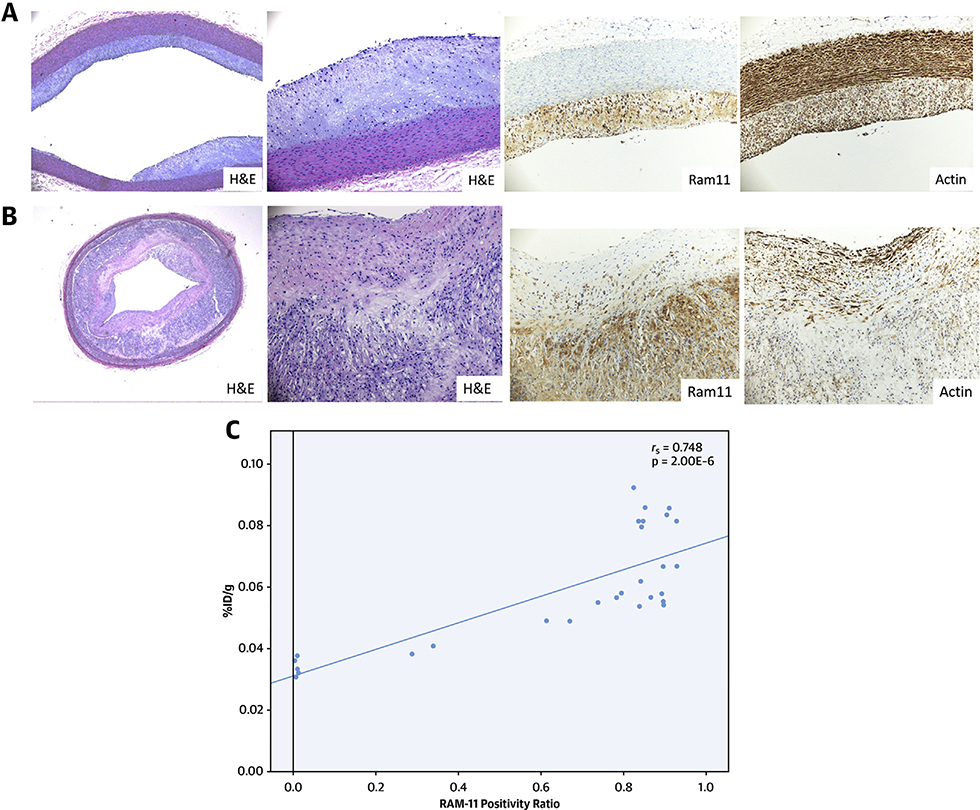
Results: A significantly higher duramycin uptake was observed in the test group compared with that of disease control and negative radiotracer control animals; duramycin uptake was also significantly higher than the annexin-V uptake. Quantitative duramycin uptake, represented as the square root of percent injected dose per cm (√ID/cm) of abdominal aorta was >2-fold higher in atherosclerotic lesions in test group (0.08 ± 0.01%) than in comparable regions of disease control animals (0.039 ± 0.0061%, p = 3.70·10-8). Mean annexin uptake (0.060 ± 0.010%) was significantly lower than duramycin (p = 0.001). Duramycin uptake corresponded to the lesion severity and macrophage burden. The radiation burden to the kidneys was substantially lower with duramycin (0.49% ID/g) than annexin (5.48% ID/g; p = 4.00·10-4).
Conclusions: Radiolabeled duramycin localizes in lipid-rich areas with high concentration of apoptotic macrophages in the experimental atherosclerosis model. Duramycin uptake in atherosclerotic lesions was significantly greater than annexin-V uptake and produced significantly lower radiation burden to nontarget organs.
Keywords: annexin-V; cell death; duramycin; radionuclide imaging; vulnerable plaques.
Copyright © 2020 American College of Cardiology Foundation. Published by Elsevier Inc. All rights reserved.
Figures
Comment in
-
Imaging Apoptosis in Atherosclerosis: From Cell Death, A Ray of Light.J Am Coll Cardiol. 2020 Oct 20;76(16):1875-1877. doi: 10.1016/j.jacc.2020.09.010. J Am Coll Cardiol. 2020. PMID: 33059833 No abstract available.
References
-
- Burke AP, Farb A, Malcom GT, Liang YH, Smialek J, Virmani R. Coronary risk factors and plaque morphology in men with coronary disease who died suddenly. N Engl J Med 1997;336: 1276–82. - PubMed
-
- Stone GW, Maehara A, Lansky AJ, et al. A prospective natural-history study of coronary atherosclerosis. N Engl J Med 2011;364:226–35. - PubMed
-
- Motoyama S, Kondo T, Sarai M, et al. Multislice computed tomographic characteristics of coronary Lesions in acute coronary syndromes. J Am Coll Cardiol 2007;50:319–26. - PubMed
Publication types
MeSH terms
Substances
Grants and funding
LinkOut - more resources
Full Text Sources
Medical
Miscellaneous
