Review
. 2020 Oct 20;76(16):1878-1901.
doi: 10.1016/j.jacc.2020.08.042.

Challenges in Cardiac and Pulmonary Sarcoidosis: JACC State-of-the-Art Review

Affiliations
Review

Challenges in Cardiac and Pulmonary Sarcoidosis: JACC State-of-the-Art Review

Maria Giovanna Trivieri et al. J Am Coll Cardiol. .

Abstract

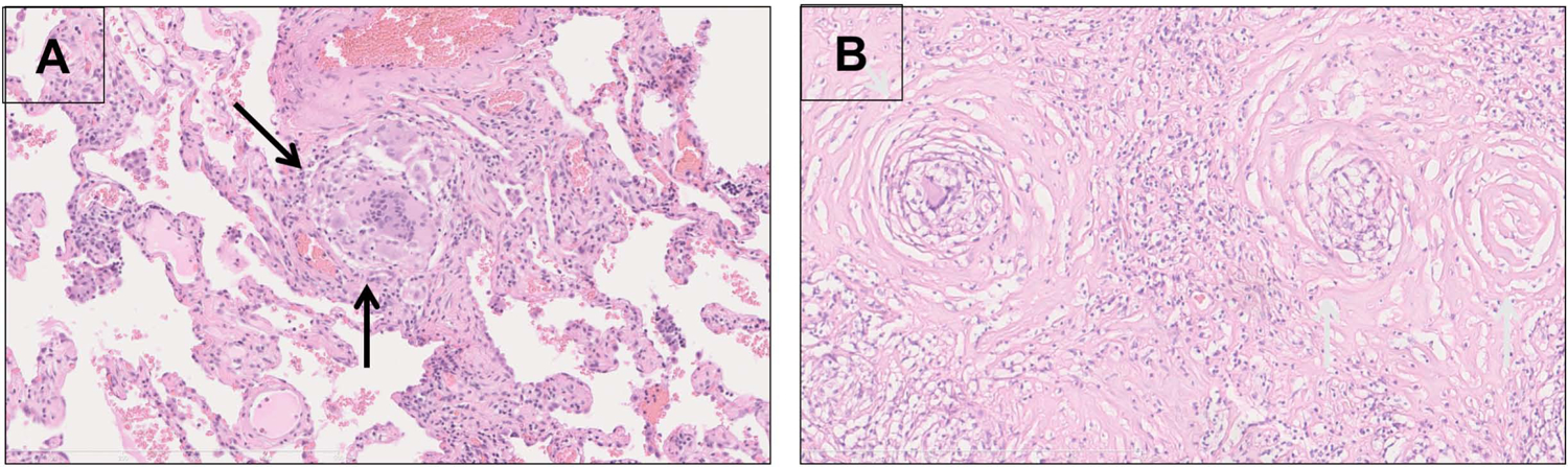
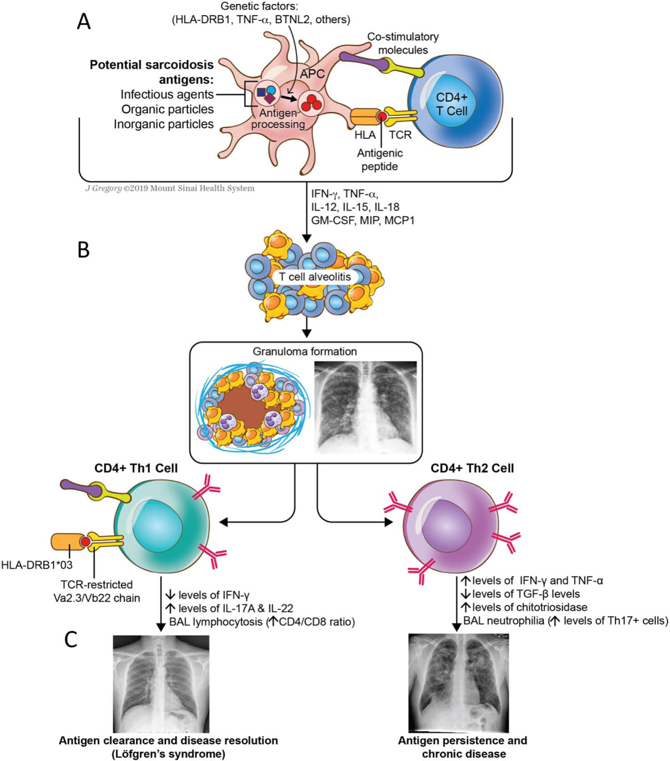
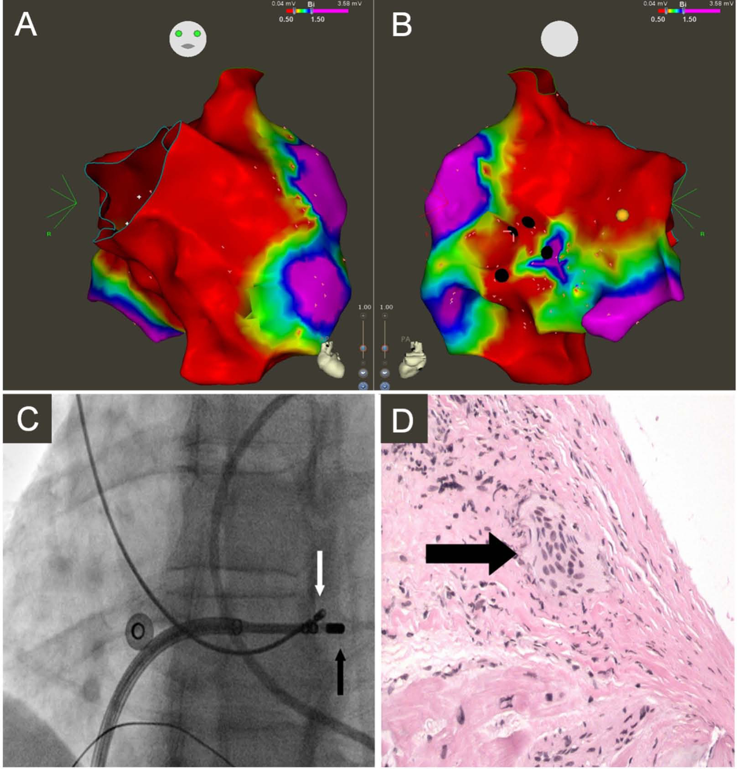
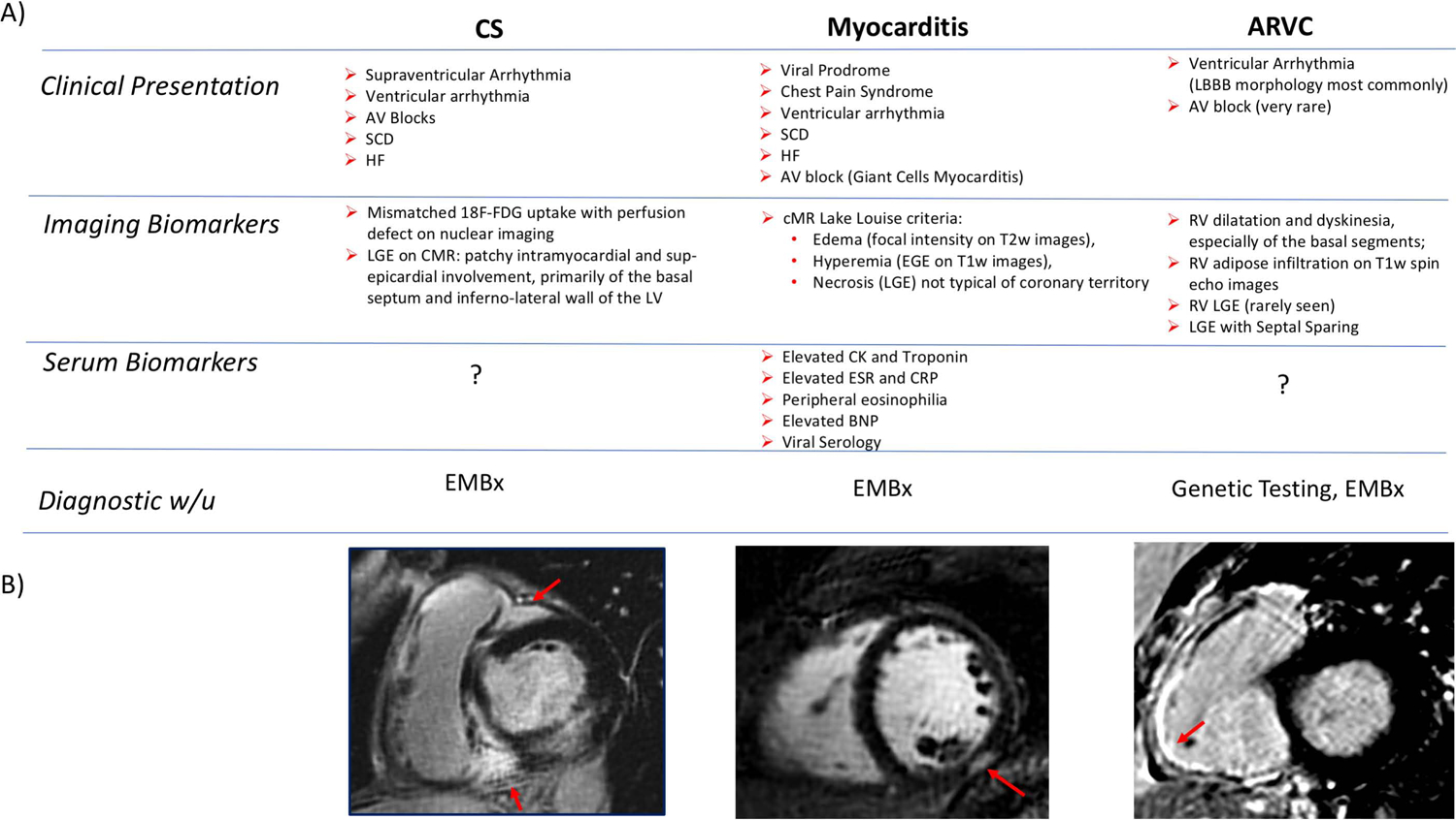
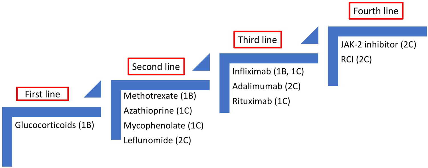
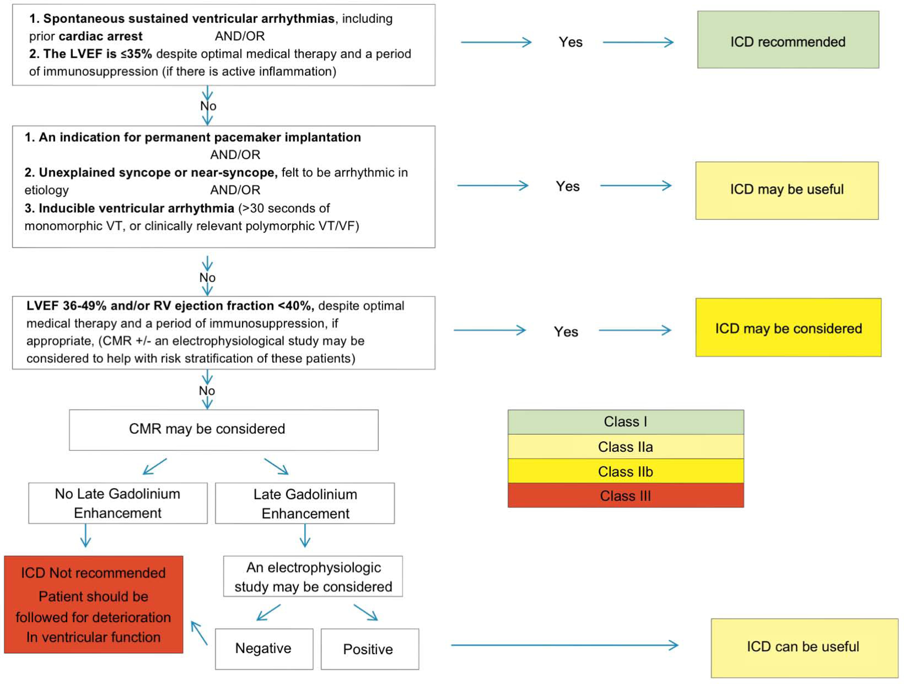
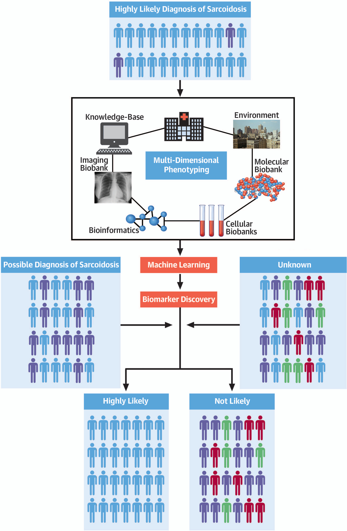
Sarcoidosis is a complex disease with heterogeneous clinical presentations that can affect virtually any organ. Although the lung is typically the most common organ involved, combined pulmonary and cardiac sarcoidosis (CS) account for most of the morbidity and mortality associated with this disease. Pulmonary sarcoidosis can be asymptomatic or result in impairment in quality of life and end-stage, severe, and/or life-threatening disease. The latter outcome is seen almost exclusively in those with fibrotic pulmonary sarcoidosis, which accounts for 10% to 20% of pulmonary sarcoidosis patients. CS is problematic to diagnose and may cause significant morbidity and death from heart failure or ventricular arrhythmias. The diagnosis of CS usually requires surrogate cardiac imaging biomarkers, as endomyocardial biopsy has relatively low yield, even with directed electrophysiological mapping. Treatment of CS is often multifactorial, involving a combination of antigranulomatous therapy and pharmacotherapy for cardiac arrhythmias and/or heart failure in addition to device placement and cardiac transplantation.

Keywords: biomarkers; cardiac sarcoidosis; imaging; pulmonary sarcoidosis.


Figures

Figure 1:
Figure 1:. Non-necrotizing Granulomas.
A: Early stage sarcoidosis (H&E stain, 20x) with a discrete perivascular granuloma (yellow arrows) comprised of epithelioid histiocytes, multinucleated giant cells, sparse lymphocytes and a lack of dense hyalinized collagen fibrosis. B: Late stage sarcoidosis (H&E stain, 20x) with a subpleural hyalinized nodule containing abundant dense eosinophilic hypocellular collagen fibrosis surrounding and bridging discrete granulomas (black arrows) with sparse lymphocytes. There prominent concentric lamellar fibrosis surrounding the granulomas, a finding characteristic of sarcoid granulomas association with scarring. Curtesy of Dr. M. A. Judson.
Figure 2:
Figure 2:. A hypothetical model of the immunopathogenesis of sarcoidosis.
Granuloma formation requires activated T-cells and macrophages coupled with a milieu of cytokines released by the immune cells. (A) T-cells are activated by a specific antigen – either environmental, infectious, or an autoantigen - presented in the context of an HLA molecule and recognized by the T-cell receptor (TCR). (B) Once activated, APCs stimulate the helper Th1-promoting cytokines such as IL-2, IL-12, IL-18, TNF-α, and IFN-γ, which orchestrate the complex process of granuloma formation and inflammation. (C) The lungs are almost universally affected by the disease. Th1 response amplification may lead to antigen clearance, disease regression (as in Löfgren’s syndrome) and remission. Failure to remove the antigen along with the involvement of a different network of cells and/or cytokines may results in chronic inflammation and fibrosis.
Figure 3:
Figure 3:. Electrophysiological-guided biopsy of the RV.
Electroanatomic bipolar voltage map of the RV displaying anterior (A) and posterior (B) views. Green, yellow, and red indicate low-voltage regions; purple denotes regions of normal voltage, defined as ≥ 1.5 mV. Black circles illustrate areas targeted for biopsy. Yellow circle illustrates location of right bundle. (C) Fluoroscopy images obtained in the left anterior oblique 25° projection showing bioptome (white arrow) targeting the low-voltage region in the RV septum, adjacent to mapping catheter (black arrow). (D) Microscopic view of an endomyocardial biopsy specimen obtained from the right ventricular septum showing noncaseating granuloma (arrow) (reproduced from(85)).
Figure 4:
Figure 4:. Diagnosis of ICS.
(A) Clinical, Imaging and Serological Biomarkers that might aid in the DDx of CS. (B) Short axis views of RV and LV showing classical patterns of LGE involving the 1) RV insertion point, basal septum as well as lateral wall in CS, 2) the sub-epicardium in myocarditis and 3) the RV free-wall in ARVC.
Figure 5:
Figure 5:. Imaging features of CS.
Short axis views of the RV/LV showing (A) areas of LGE in the RV, basal septum as well as RV lateral wall with matching increased 18F–FDG signal (B, C) perfectly co-localized to area of LGE (MR+PET+) on fused 18F–FDG-PET/MR images. This pattern of LGE and 18F–FDG uptake (“hug sign”) was solely observed in patients with biopsy proven CS and might constitute the imaging biomarker “signature” of the disease. This pattern faithfully replicated the one previously identified in explanted hearts from patients with ICS (D). Reproduced with permission (62).
Figure 6:
Figure 6:. Proposed diagnostic and management approach for the assessment and treatment of CS.
The treatment of CS can be divided into two general categories: treatment of the inflammation or of its clinical sequelae, i.e. arrhythmias and ventricular dysfunction. The figure summarizes the available imaging modalities, diagnostic and therapeutic options.
Figure 7:
Figure 7:. PET scan as a possible tool to predict response to anti-inflammatory therapy and to guide treatment decision.
Cardiac PET/MR was performed in a patient with biopsy proven CS symptomatic with AF. The initial scan revealed 18F–FDG avid mediastinal LNDs as well as cardiac uptake in the basal segments of the antero-septum. IS therapy with steroids was instituted, with spontaneous cardioversion. Follow up scan at 3 and 6 months revealed progressive resolution of the inflammation.
Figure 8:
Figure 8:. Suggested treatment algorithm for patients with clinically manifest CS. The figure proposes an imaging guided approach to escalation and de-escalation of immunosuppressive therapy.
Reproduced with permission (5).
Figure 9:
Figure 9:. Stepwise approach to the therapy for CS.
Treatments were scored based on reported effectiveness for cardiac (1) or extra-cardiac (2) sarcoidosis. Level of evidence: A for randomized trials, B for case series or C for case reports. There is presently no randomized trial in CS. Initiation and escalation of treatment is commonly guided by PET.
Figure 10:
Figure 10:. ICD Implantation for CS.
Consensus recommendations for ICD implantation in patients diagnosed with Cardiac Sarcoidosis. Reproduced from (89)
Central Illustration:
Central Illustration:. Precision Sarcoidosis.
The integration of clinical, biological, omics and imaging data point from highly selected cases of sarcoidosis coupled with a standardized approach to patient phenotyping and machine learning algorithms is a promising pathway to enable the discovery of multidimensional disease specific biomarkers to use for accurate patient selection and targeted treatments.

Similar articles

Cited by

References

    1. Arkema EV, Cozier YC. Epidemiology of sarcoidosis: current findings and future directions. Ther Adv Chronic Dis 2018;9:227–240. - PMC - PubMed
    1. Judson MA, Boan AD, Lackland DT. The clinical course of sarcoidosis: presentation, diagnosis, and treatment in a large white and black cohort in the United States. Sarcoidosis Vasc Diffuse Lung Dis 2012;29:119–27. - PubMed
    1. Morimoto T, Azuma A et al. Epidemiology of sarcoidosis in Japan. Eur Respir J 2008;31:372–9. - PubMed
    1. Ohta H, Tazawa R et al. Acute-onset sarcoidosis with erythema nodosum and polyarthralgia (Lofgren’s syndrome) in Japan: a case report and a review of the literature. Intern Med 2006;45:659–62. - PubMed
    1. Birnie DH, Nery PB et al. Cardiac Sarcoidosis. J Am Coll Cardiol 2016;68:411–21. - PubMed

Publication types

MeSH terms

Substances