

Centrally Reduced Diffusion Sign for Differentiation between Treatment-Related Lesions and Glioma Progression: A Validation Study
- PMID: 33060101
- PMCID: PMC7658838
- DOI: 10.3174/ajnr.A6843
Centrally Reduced Diffusion Sign for Differentiation between Treatment-Related Lesions and Glioma Progression: A Validation Study
Abstract
Background and purpose: Differentiating between treatment-related lesions and tumor progression remains one of the greatest dilemmas in neuro-oncology. Diffusion MR imaging characteristics may provide useful information to help make this distinction. The aim of the study was to assess the diagnostic accuracy of the centrally reduced diffusion sign for differentiation of treatment-related lesions and true tumor progression in patients with suspected glioma recurrence.
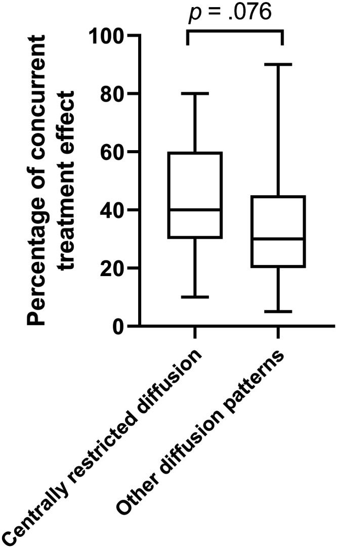
Materials and methods: The images of 231 patients who underwent an operation for suspected glioma recurrence were reviewed. Patients with susceptibility artifacts or without central necrosis were excluded. The final diagnosis was established according to histopathology reports. Two neuroradiologists classified the diffusion patterns on preoperative MR imaging as the following: 1) reduced diffusion in the solid component only, 2) reduced diffusion mainly in the solid component, 3) no reduced diffusion, 4) reduced diffusion mainly in the central necrosis, and 5) reduced diffusion in the central necrosis only. Diagnostic accuracy metrics and the area under the receiver operating characteristic curve were estimated for the diffusion patterns.
Results: One hundred three patients were included (22 with treatment-related lesions and 81 with tumor progression). The diagnostic accuracy results for the centrally reduced diffusion pattern as a predictor of treatment-related lesions ("mainly central" and "exclusively central" patterns versus all other patterns) were as follows: 64% sensitivity (95% CI, 41%-83%), 84% specificity (95% CI, 74%-91%), 52% positive predictive value (95% CI, 37%-66%), and 89% negative predictive value (95% CI, 83%-94%).
Conclusions: The centrally reduced diffusion sign is associated with the presence of treatment effect. The probability of a histologic diagnosis of a treatment-related lesion is low (11%) in the absence of centrally reduced diffusion.
© 2020 by American Journal of Neuroradiology.
Figures


References
-
- Ceschin R, Kurland BF, Abberbock SR, et al. Parametric response mapping of apparent diffusion coefficient as an imaging biomarker to distinguish pseudoprogression from true tumor progression in peptide-based vaccine therapy for pediatric diffuse intrinsic pontine glioma. AJNR Am J Neuroradiol 2015;36:2170–76 10.3174/ajnr.A4428 - DOI - PMC - PubMed