. 2021 Feb 1;27(3):775-784.
doi: 10.1158/1078-0432.CCR-20-0894. Epub 2020 Oct 15.

Hypofractionated FLASH-RT as an Effective Treatment against Glioblastoma that Reduces Neurocognitive Side Effects in Mice

Affiliations

Hypofractionated FLASH-RT as an Effective Treatment against Glioblastoma that Reduces Neurocognitive Side Effects in Mice

Pierre Montay-Gruel et al. Clin Cancer Res. .

Abstract

Purpose: Recent data have shown that single-fraction irradiation delivered to the whole brain in less than tenths of a second using FLASH radiotherapy (FLASH-RT), does not elicit neurocognitive deficits in mice. This observation has important clinical implications for the management of invasive and treatment-resistant brain tumors that involves relatively large irradiation volumes with high cytotoxic doses.

Experimental design: Therefore, we aimed at simultaneously investigating the antitumor efficacy and neuroprotective benefits of FLASH-RT 1-month after exposure, using a well-characterized murine orthotopic glioblastoma model. As fractionated regimens of radiotherapy are the standard of care for glioblastoma treatment, we incorporated dose fractionation to simultaneously validate the neuroprotective effects and optimized tumor treatments with FLASH-RT.

Results: The capability of FLASH-RT to minimize the induction of radiation-induced brain toxicities has been attributed to the reduction of reactive oxygen species, casting some concern that this might translate to a possible loss of antitumor efficacy. Our study shows that FLASH and CONV-RT are isoefficient in delaying glioblastoma growth for all tested regimens. Furthermore, only FLASH-RT was found to significantly spare radiation-induced cognitive deficits in learning and memory in tumor-bearing animals after the delivery of large neurotoxic single dose or hypofractionated regimens.

Conclusions: The present results show that FLASH-RT delivered with hypofractionated regimens is able to spare the normal brain from radiation-induced toxicities without compromising tumor cure. This exciting capability provides an initial framework for future clinical applications of FLASH-RT.See related commentary by Huang and Mendonca, p. 662.


Conflict of interest statement

Conflict of interest

The authors declare no conflicts of interest.

Figures

Figure 1:
Figure 1:
Summary of the temporal dosimetry characteristics of the published experimental data describing the FLASH effect in vivo (–,,,,–37) or in vitro (–42) (colored dots), those that have not been able to observe the FLASH effect (43,44) (black, grey) and the dose rate de-escalation studies showing the range in which the FLASH effect is lost (colored crosses). The horizontal axis denotes the dose rate per pulse for electrons (e) and protons (p) or in a single stripe (as described in (27)) for synchrotron radiation (Rx). The vertical axis corresponds to the total irradiation time needed for delivering 10 Gy at the average dose rate quoted by the authors of the publications. Parameters for other dose values have been changed accordingly. Adapted from Bourhis et al., 2019 (15).
Figure 2:
Figure 2:
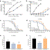
Tumor growth delay of H454 orthotopic GBM implanted in the striatum of female Nude mice measured by bioluminescence (a, b, c) treated with 0, 10 Gy (BED = 20 Gy), 14 Gy (BED = 33.6 Gy) single dose or 2 ☓ 7 Gy (BED = 23.8 Gy) daily fractionated WBI delivered with FLASH or CONV-RT. Mean change in relative bioluminescence ± SEM, N = 10–12 animals per group. P values were derived from the Mann–Whitney U test: **P < 0.01; ***P < 0.001 compared FLASH vs CONV group; ns: not significant. α/β ratio of 10 for BED calculation on the tumor. Survival curves of glioblastoma bearing mice treated with 0, 10 or 14 Gy single dose or 2 ☓ 7 Gy daily fractionated WBI with FLASH or CONV-RT (d, e, f). N = 10–12 animals per group. P values were derived from the log-rank test; compared FLASH vs CONV group ns: not significant. Memory skills of glioblastoma bearing mice treated with 0, 10 Gy (BED = 43.3 Gy), 14 Gy (BED = 79.3 Gy) single dose or 2 ☓ 7 Gy (BED = 46.7 Gy) daily fractionated WBI delivered with FLASH or CONV-RT, evaluated by Novel Object Recognition test 4 weeks post-implantation (g, h, i). Mean DI ± SEM, N = 10–14 animals per group. P values were derived from the Mann–Whitney U test: *P < 0.05; **P < 0.01 compared Control and FLASH vs CONV group. ns: not significant. α/β ratio of 3 for BED calculation on the normal brain tissue.
Figure 3:
Figure 3:
Tumor growth delay of H454 orthotopic GBM implanted in the striatum of female Nude mice treated with 0, 4 ☓ 3.5 Gy (BED = 18.9 Gy) daily fractionated WBI; or 3 ☓ 10 Gy (BED = 60 Gy) spaced by 48h WBI delivered with FLASH or CONV-RT (a, b). Mean change in relative bioluminescence ± SEM, N = 9–13 animals per group. P values were derived from the Mann–Whitney U test: **P < 0.01; ***P < 0.001; ****P < 0.0001 compared FLASH vs CONV group; ns: not significant. α/β ratio of 10 for BED calculation on the tumor. Survival curves of glioblastoma bearing mice treated with 0, 4 ☓ 3.5 daily fractionated WBI; or 3 ☓ 10 Gy spaced by 48h WBI delivered with FLASH or CONV-RT (c, d). N = 9–13 animals per group. P values were derived from the log-rank test. ***P < 0.001; ****P < 0.0001 vs. control group; ns: not significant. Memory skills of glioblastoma bearing mice treated with 0 Gy, 4 ☓ 3.5 Gy (BED = 30.3 Gy) daily fractionated WBI; or 3 ☓ 10 Gy (BED = 130 Gy) spaced by 48h WBI delivered with FLASH or CONV-RT, evaluated by Novel Object Recognition test 4 weeks post-implantation (e, f). Mean DI ± SEM, N = 8–12 animals per group. P values were derived from the Mann–Whitney U test: *P < 0.05; **P < 0.01 compared with the CONV group. ns: not significant. α/β ratio of 3 for BED calculation on the normal brain tissue.
Figure 4:
Figure 4:
Tumor growth delay of H454 orthotopic GBM implanted in the striatum of female Nude mice measured by bioluminescence (a) treated with 25 Gy (BED = 233 Gy) single dose HBI delivered with FLASH or CONV-RT. Mean change in relative bioluminescence ± SEM, N = 9–10 animals per group. P values were derived from the Mann–Whitney U test: ****P < 0.0001; ns: not significant. α/β ratio of 10 for BED calculation on the tumor. Survival curves of H454 GBM bearing mice (b) treated with 25 Gy single dose HBI delivered with FLASH or CONV-RT. N = 10 animals per group. P values were derived from log-rank test: ***P < 0.01; ****P < 0.0001 compared with the control group. ns: not significant. Memory skills of glioblastoma bearing mice treated with 25 Gy (BED = 87.5 Gy) single dose HBI delivered with FLASH or CONV-RT, evaluated by Novel Object Recognition test 4 weeks post-implantation (c). Mean ± SEM, N = 9–10 animals per group. P values were derived from the Mann–Whitney U test: ns: not significant. α/β ratio of 3 for BED calculation on the normal brain tissue.
Figure 5:
Figure 5:
Relative tumor growth delay of H454 orthotopic GBM as a function of the Biologically Effective Dose (BED) delivered to the tumor with FLASH or CONV-RT, 3 weeks post-irradiation (a). BED on the tumor was calculated with the following formula: nd×(1+dα/β), where n is the number of fractions, d is the dose per fraction and the α/β ratio is set to 10 (b).

Comment in

Similar articles

Cited by

References

    1. Delaney G, Jacob S, Featherstone C, Barton M. The role of radiotherapy in cancer treatment: Estimating optimal utilization from a review of evidence-based clinical guidelines. Cancer. 2005;104:1129–37. - PubMed
    1. Rosenblatt E, Izewska J, Anacak Y, Pynda Y, Scalliet P, Boniol M, et al. Radiotherapy capacity in European countries: An analysis of the Directory of Radiotherapy Centres (DIRAC) database. Lancet Oncol. 2013;14. - PubMed
    1. Meyers CA, Hess KR, Yung WKA, Levin VA. Cognitive function as a predictor of survival in patients with recurrent malignant glioma. J Clin Oncol. 2000;18:646–50. - PubMed
    1. Butler JM, Rapp SR, Shaw EG. Managing the cognitive effects of brain tumor radiation therapy. Curr Treat Options Oncol. 2006;7:517–23. - PubMed
    1. Wellisch DK, Kaleita TA, Freeman D, Cloughesy T, Goldman J. Predicting major depression in brain tumor patients. Psychooncology. 2002;11:230–8. - PubMed

Publication types

MeSH terms

Substances