. 2020 Nov 27;147(22):dev193862.
doi: 10.1242/dev.193862.

The conserved molting/circadian rhythm regulator NHR-23/NR1F1 serves as an essential co-regulator of C. elegans spermatogenesis

Affiliations

The conserved molting/circadian rhythm regulator NHR-23/NR1F1 serves as an essential co-regulator of C. elegans spermatogenesis

James Matthew Ragle et al. Development. .

Abstract

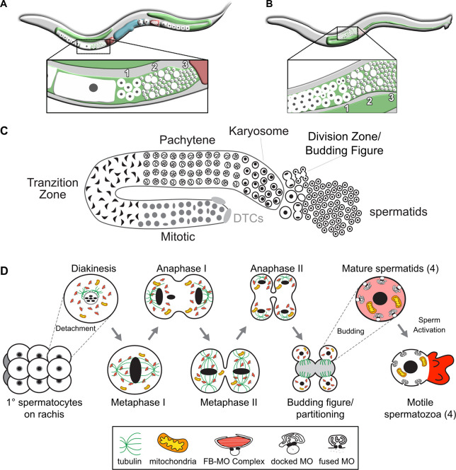
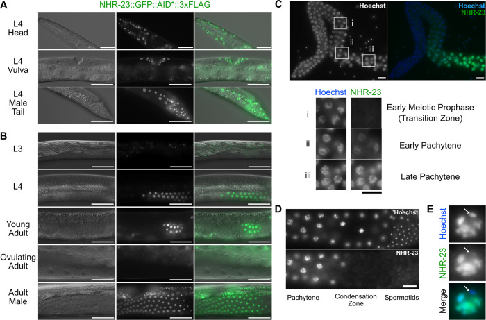
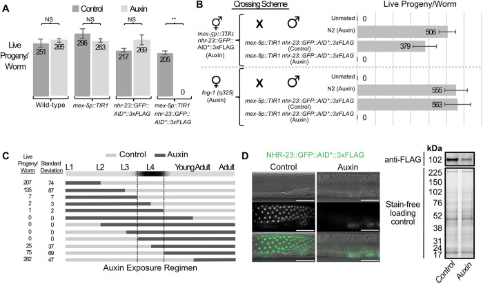
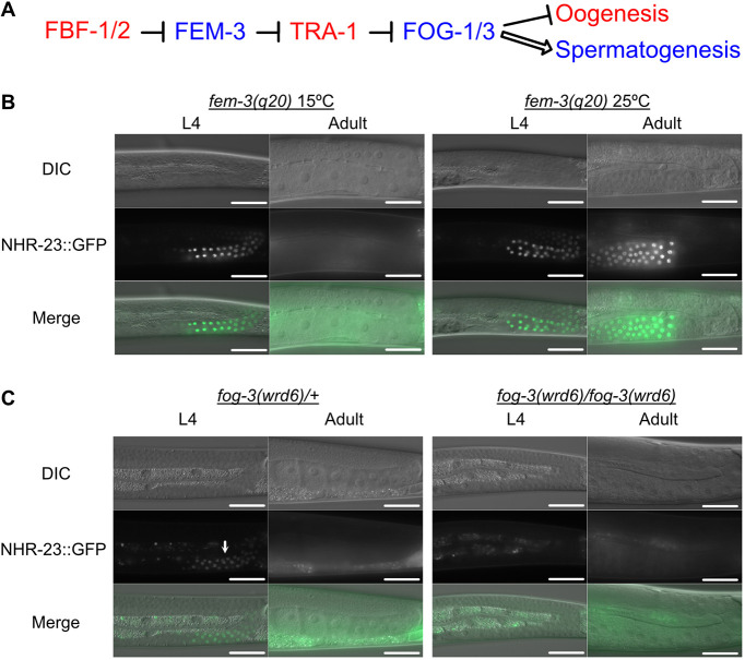
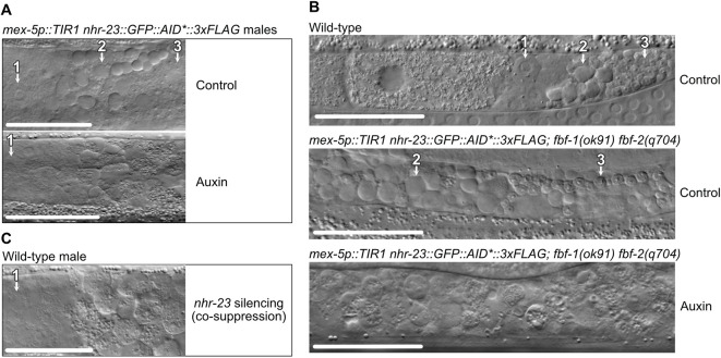
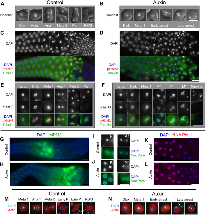
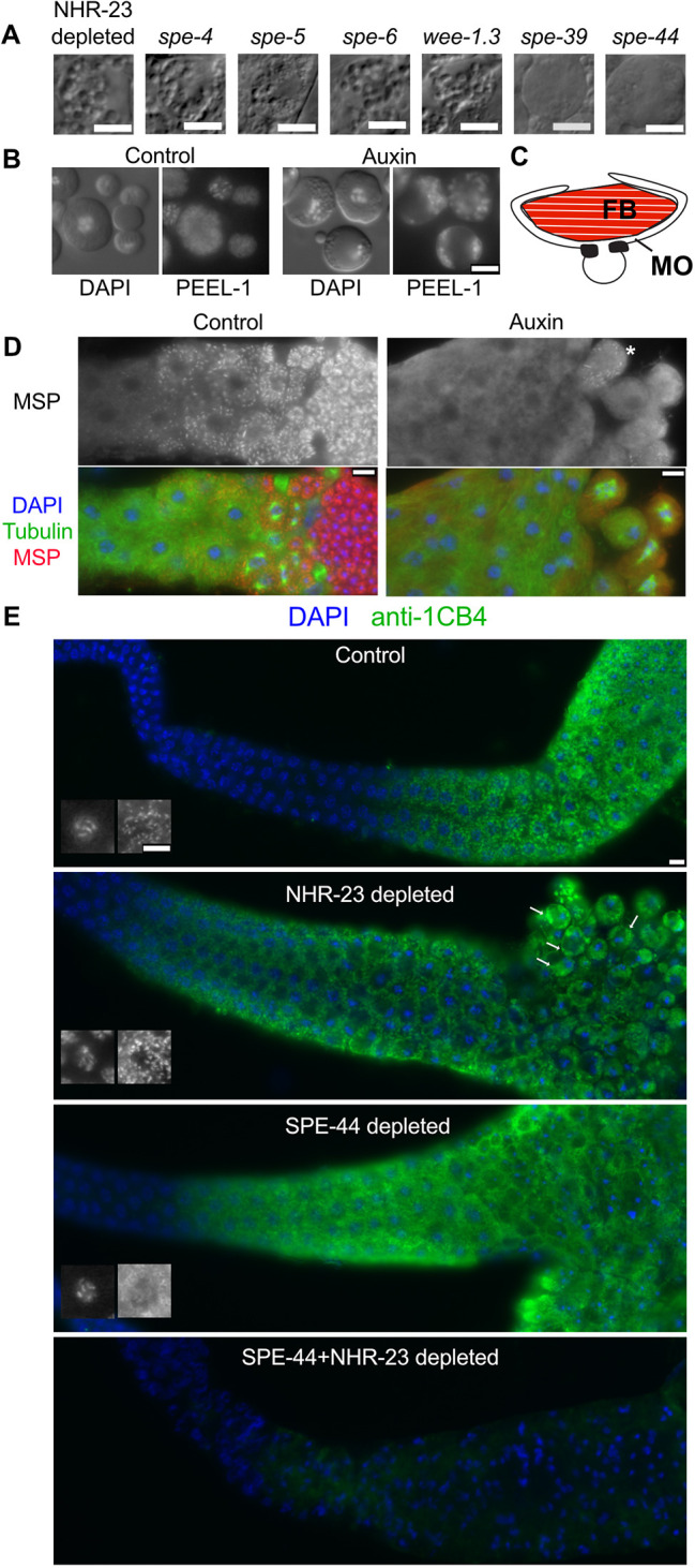
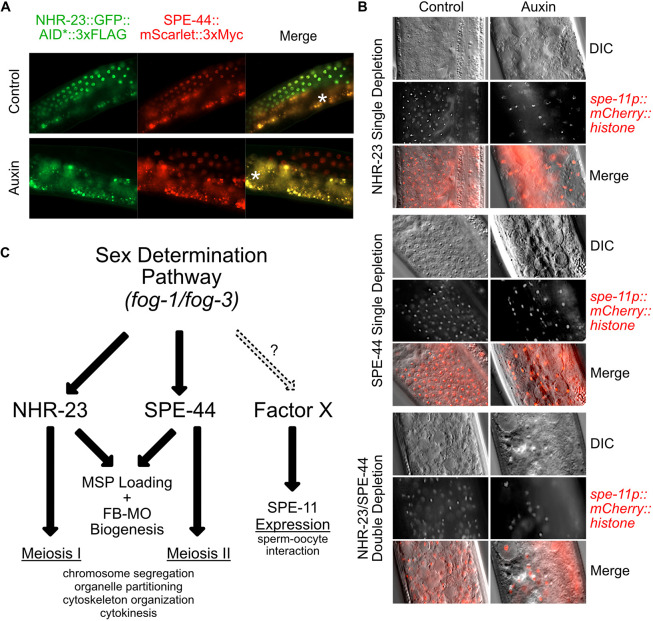
In sexually reproducing metazoans, spermatogenesis is the process by which uncommitted germ cells give rise to haploid sperm. Work in model systems has revealed mechanisms controlling commitment to the sperm fate, but how this fate is subsequently executed remains less clear. While studying the well-established role of the conserved nuclear hormone receptor transcription factor, NHR-23/NR1F1, in regulating C. elegans molting, we discovered that NHR-23/NR1F1 is also constitutively expressed in developing primary spermatocytes and is a critical regulator of spermatogenesis. In this novel role, NHR-23/NR1F1 functions downstream of the canonical sex-determination pathway. Degron-mediated depletion of NHR-23/NR1F1 within hermaphrodite or male germlines causes sterility due to an absence of functional sperm, as depleted animals produce arrested primary spermatocytes rather than haploid sperm. These spermatocytes arrest in prometaphase I and fail to either progress to anaphase or attempt spermatid-residual body partitioning. They make sperm-specific membranous organelles but fail to assemble their major sperm protein into fibrous bodies. NHR-23/NR1F1 appears to function independently of the known SPE-44 gene regulatory network, revealing the existence of an NHR-23/NR1F1-mediated module that regulates the spermatogenesis program.

Keywords: Auxin-inducible degron; C. elegans; Meiosis; NHR-23; Nuclear hormone receptor; Spermatogenesis.


Conflict of interest statement

Competing interestsThe authors declare no competing or financial interests.

Figures

Fig. 1.
Fig. 1.
Overview of C. elegans spermatogenesis. (A,B) Cartoons depicting a young adult C. elegans hermaphrodite (A) and male (B), and their respective germlines. The hermaphrodite germline (A) is transitioning from spermatogenesis to oogenesis. The enlarged views highlight the linear arrangement of the primary spermatocytes (1), residual bodies (RBs) (2) and mature haploid spermatids (3). (C) Stylized cartoon of a surface view of the male germline highlighting its overall linear organization. Mitotic proliferation of the germline stem cells is maintained by two somatic distal tip cells (DTCs) that form the germ cell niche. The early events of meiotic prophase, including homolog pairing and formation of the synaptonemal complex, occur in the transition zone. Following an extended pachytene stage, spermatocytes enter a karyosome stage before mature spermatocytes detach from the syncytial germline and divide meiotically. The first meiotic division is often incomplete, leaving secondary spermatocytes linked by a cytoplasmic connection. Following anaphase II, the spermatocytes morph into budding figures that split into residual bodies and haploid spermatids. (D) Details of the meiotic divisions and post-meiotic partitioning event. Once spermatocytes detach from the germline syncytium, they pass through a brief diakinesis stage before undergoing nuclear envelope breakdown and initiating meiotic divisions. During the post-meiotic partitioning event, microtubules become acentrosomal and localize to the developing residual body (Winter et al., 2017). Components retained in the spermatids include fibrous body-membranous organelles (FB-MO), mitochondria, chromatin and centrioles. Components discarded within the RB include the tubulin, actin, endoplasmic reticulum and ribosomes of the cell; mature sperm are thus both transcriptionally and translationally inactive. Following separation from the RB, FBs disassemble and release unpolymerized MSP and the MOs dock with the plasma membrane. Males store sperm in this inactive spermatid state. During spermatid activation, MOs fuse with the plasma membrane and unpolymerized MSP localizes to the pseudopod, where it forms fibers that are required for spermatozoon motility.
Fig. 2.
Fig. 2.
NHR-23::GFP::AID*::3xFLAG is expressed in somatic cells throughout development and in spermatogenic germlines. A strain carrying GFP::AID*::3xFLAG knocked-in to the endogenous nhr-23 gene to produce a C-terminal translational fusion to all known nhr-23 isoforms was used to monitor endogenous NHR-23 expression. (A) Representative DIC and GFP images of NHR-23::GFP::AID*::3xFLAG L4 larvae, specifically hypodermal cells of a hermaphrodite head, vulval precursor cells and seam/hypodermal cells of the male tail. (B) Representative DIC and GFP images of NHR-23::GFP::AID*::3xFLAG in pachytene cells of the germline in L3, L4, young adult, ovulating adult and adult male worms. (C) Fluorescent images of NHR-23::GFP::AID*::3xFLAG in a dissected adult male germline. (i-iii) Cells in early meiotic prophase (transition zone) (i), and in early (ii) and late (iii) pachytene. (D) Representative fluorescent images of NHR-23::GFP::AID*::3xFLAG in a late pachytene male germline. (E) Representative image of a late pachytene nucleus expressing NHR-23::GFP::AID*::3xFLAG. The location of the X chromosome is indicated with a white arrow. Scale bars: 40 µm in A,B; 10 µm in C-E. A minimum of 12 P0 animals were analyzed in A-C. Nuclei are visualized using Hoechst stain in C-E.
Fig. 3.
Fig. 3.
NHR-23-depletion in the germline during periods of spermatogenesis causes infertility in hermaphrodites and males. (A) Average number of live progeny produced by wild-type, mex-5p::TIR1, nhr-23::GFP::AID*::3xFLAG or mex-5p::TIR1 nhr-23::GFP::AID*::3xFLAG hermaphrodites grown from L1 through adulthood on MYOB media with or without 4 mM auxin. Student's t-test was performed for each genotype comparing brood sizes on control media versus 4 mM auxin. P>0.01 was considered non-significant (NS). **P<0.00001. n=12 for each brood size. Data are mean±s.e.m. (B) Average number of live progeny produced by mex-5p::TIR1 nhr-23::GFP::AID*::3xFLAG or fog-1(q325) hermaphrodites grown from L1 on MYOB media with 4 mM auxin. Hermaphrodites were either unmated or crossed to males of the indicated genotype; males were grown from L1 to adulthood on control or 4 mM auxin media (n=12). Data are mean±s.e.m. (C) Average number of live progeny produced by mex-5p::TIR1 nhr-23::GFP::AID*::3xFLAG hermaphrodites shifted on or off 4 mM auxin at different points in development. Dark horizontal bands represent growth on media with 4 mM auxin and light horizontal bands represent growth on control media lacking auxin. n=12 for each condition. (D) DIC and fluorescent images of mex-5p::TIR1 nhr-23::GFP::AID*::3xFLAG adult male germlines after animals were grown from L1 on MYOB media±4 mM auxin. NHR-23-depletion results in minimally detectable GFP signal by fluorescence microscopy, but FLAG signal remains detectable in western blots. Scale bars: 40 μm. Anti-FLAG immunoblot analyses of lysates are from synchronized male mex-5p::TIR1 nhr-23::GFP::AID*::3xFLAG animals grown on control or 4 mM auxin media. Marker size (in kDa) is provided. Predicted size of the fusion protein is 98.7 kDa, based on the NHR-23 isoform expressed in adults in a Nanopore direct RNA-sequencing dataset (Roach et al., 2020). Stain-free analysis, which visualizes total protein on the membrane is provided as a loading control.
Fig. 4.
Fig. 4.
nhr-23 is downstream of the germline sex-determination pathway. (A) Simplified schematic of the C. elegans sex-determination pathway and their effect on gamete fate. The wild-type function of the factors in blue is to promote spermatogenesis, whereas the factors in red promote oogenesis. (B) DIC and GFP images of mex-5p::TIR1 nhr-23::GFP::AID*:: 3xFLAG; fem-3(q20) L4 and adult hermaphrodites grown from L1 onwards at permissive (15°C) or restrictive (25°C) temperatures. (C) DIC and GFP images of mex-5p::TIR1 nhr-23::GFP::AID*::3xFLAG; fog-3(wrd6) L4 and adult hermaphrodites. The white arrow indicates NHR-23::GFP::AID*::3xFLAG expression in late pachytene cells of a fog-3(wrd6) heterozygous hermaphrodite germline. Scale bars: 40 µm.
Fig. 5.
Fig. 5.
NHR-23 is necessary for sperm development. (A) DIC images of the proximal germlines of mex-5p::TIR1, nhr-23::GFP::AID*::3xFLAG young adult males grown from L1 onwards on control or 4 mM auxin media. (B) DIC images of wild-type and masculinized mex-5p::TIR1, nhr-23::GFP::AID*::3xFLAG; fbf-1(ok91) fbf-2(q704) hermaphrodites grown from L1 onwards on control or 4 mM auxin media. (1) Primary spermatocytes have a flat morphology with a large nucleus. (2) Residual bodies appear as highly refractile raised button-like structures. (3) Spermatids are readily discernible through their characteristic small refractive nuclei. (C) DIC image of the proximal germline in a wild-type young adult male F1 progeny from a P0 animal injected with a mex-5p::nhr-23 cDNA co-suppression construct, which promotes an RNAi-like depletion of targeted mRNA. Scale bars: 40 µm.
Fig. 6.
Fig. 6.
Metaphase I-like arrest in NHR-23-depleted spermatocytes. (A,B) Live spermatocytes ordered according to stage. Differential interference contrast (DIC) images of cells were overlaid by epifluorescence images of their Hoechst stained nuclei. (C-J) Isolated and fixed male gonads, and individual spermatocytes labeled with DAPI (blue) and indicated antibodies. (C,D) Gonad images show the proximal gonad from late pachytene (P) to either haploid spermatids (S) or terminal arrest (T) co-labeled with antibodies against α-tubulin (green) and phosphorylated histone H3 (ser10) (red). Controls (nhr-23::AID* without auxin or him-5 with auxin) in C; nhr-23::AID* with auxin in D. (E,F) Higher magnification images of individual spermatocytes. (G,H) Isolated control (G) and NHR-23-depleted (H) proximal male gonads (pachytene and later meiotic stages) co-labeled with antibodies against MPM2 (green), which binds diverse mitotic and meiotic phosphorylated proteins. (I,J) Staged spermatocytes co-labeled with anti-nuclear pore protein (green) in control (I) and NHR-23-depleted (J) males. (K,L) Control (K) and NHR-23-depleted (L) gonads (pachytene through karyosome) co-labeled with anti-phospho-RNA polymerase II CTD repeat (red) show switching off of global transcription in karyosome spermatocytes. (M,N) Aldehyde-fixed and staged spermatocytes with DNA pseudo-colored in cyan (DAPI) and actin microfilaments labeled with rhodamine. Arrow in N indicates the chromatin of an adjacent lysed cell. P, pachytene; K, karyosome; Div, meiotic divisions; Diak, diakinesis; Meta 1/M1, metaphase I; M*, aberrant metaphase I; Ana1/A1, anaphase I; Meta 2, metaphase II; Par, post-meiotic partitioning; RB, residual body; S, haploid spermatids; T, terminal-stage spermatocyte. Scale bars: 10 µm.
Fig. 7.
Fig. 7.
FB-MO defects in NHR-23 spermatocytes and synergy with SPE-44. (A) DIC images of arrested spermatocytes from spermatogenesis-defective mutants. (B) Control and NHR-23-depleted spermatocytes visualized by DIC/Hoechst (left) and the MO marker PEEL::GFP (right). (C) Schematic of an FB-MO complex showing an MSP-enriched fibrous body (FB) enveloped within arm-like extensions of the Golgi-derived membranous organelle (MO). (D) Proximal gonads from control (left) and NHR-23-depleted (right) males co-labeled with DAPI and antibodies against tubulin and MSP. Asterisk indicates a single spermatocyte in the NHR-23-depleted gonad with MSP polymers. (E) Gonads from animals with indicated protein depletions co-labeled with DAPI and the anti-MO antibody 1CB4. Black and white insets show DAPI staining (left) and 1CB4-stained MOs (right) in pachytene stage spermatocytes and highlight defects in SPE-44 spermatocytes. Arrows indicate clumping of 1CB4-stained bodies following NHR-23 depletion. Scale bars: 5 µm.
Fig. 8.
Fig. 8.
Control of transcription in spermatogenesis is facilitated by multiple factors with divergent targets. (A) Fluorescent images of NHR-23::GFP::AID*::3xFLAG and spe-44::mScarlet::3xMyc. Worms containing both translational fusions as well as the mex-5p::TIR1 driver were grown from L1 onwards on control or 4 mM auxin media. The asterisks indicate gut autofluorescence. (B) Representative DIC and fluorescent images of spe-11p::mCherry::H2B in adult males expressing mex-5p::TIR1 in the germline. Worms individually expressed NHR-23::AID*::3xFLAG or SPE-44::AID*::3xFLAG, or both, were grown from L1 until the young adult stage on control or 4 mM auxin media. (C) Model depicting the coordinated control of gene expression prior to and during the stages of spermatogenesis. NHR-23 and SPE-44 control distinct sets of genes to promote the events of meiosis I and II, respectively. They redundantly promote MSP loading and FB-MO biogenesis. A third pathway controlled by yet unidentified factors promotes the expression of genes involved in the sperm-oocyte interaction, such as spe-11.

Similar articles

Cited by

References

    1. Akashi M. and Takumi T. (2005). The orphan nuclear receptor RORα regulates circadian transcription of the mammalian core-clock Bmal1. Nat. Struct. Mol. Biol. 12, 441-448. 10.1038/nsmb925 - DOI - PubMed
    1. Alvarez J. D., Hansen A., Ord T., Bebas P., Chappell P. E., Giebultowicz J. M., Williams C., Moss S. and Sehgal A. (2008). The circadian clock protein BMAL1 is necessary for fertility and proper testosterone production in mice. J. Biol. Rhythms 23, 26-36. 10.1177/0748730407311254 - DOI - PMC - PubMed
    1. André E., Conquet F., Steinmayr M., Stratton S. C., Porciatti V. and Becker-André M. (1998). Disruption of retinoid-related orphan receptor β changes circadian behavior, causes retinal degeneration and leads to vacillans phenotype in mice. EMBO J. 17, 3867-3877. 10.1093/emboj/17.14.3867 - DOI - PMC - PubMed
    1. Arribere J. A., Bell R. T., Fu B. X. H., Artiles K. L., Hartman P. S. and Fire A. Z. (2014). Efficient marker-free recovery of custom genetic modifications with CRISPR/Cas9 in Caenorhabditis elegans. Genetics 198, 837-846. 10.1534/genetics.114.169730 - DOI - PMC - PubMed
    1. Ashley G., Duong T., Levenson M. T., Martinez M. A. Q., Hibshman J. D., Saeger H. N., Doonan R., Palmisano N. J., Martinez-Mendez R., Davidson B. et al. (2020). Expanding the Caenorhabditis elegans auxin-inducible degron system toolkit with internal expression and degradation controls and improved modular constructs for CRISPR/Cas9-mediated genome editing. bioRxiv. 10.1101/2020.05.12.090217 - DOI

Publication types

MeSH terms

Substances