. 2020 Nov 1;34(21-22):1452-1473.
doi: 10.1101/gad.341545.120. Epub 2020 Oct 15.

Selective inhibition of CDK7 reveals high-confidence targets and new models for TFIIH function in transcription

Affiliations

Selective inhibition of CDK7 reveals high-confidence targets and new models for TFIIH function in transcription

Jenna K Rimel et al. Genes Dev. .

Abstract

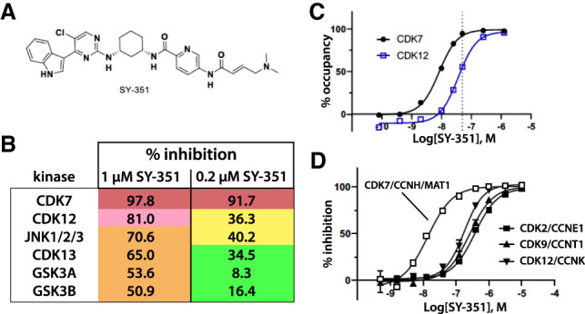
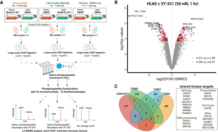
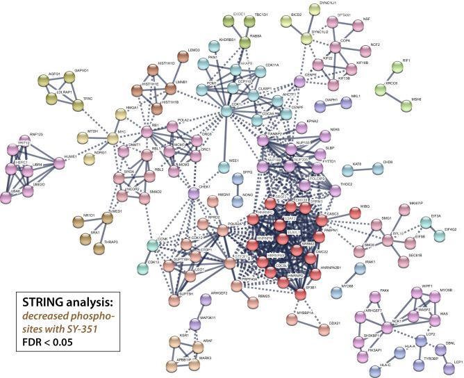
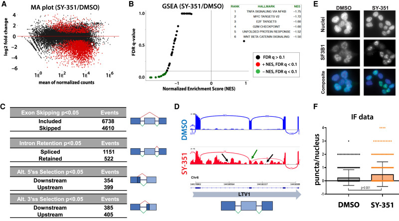
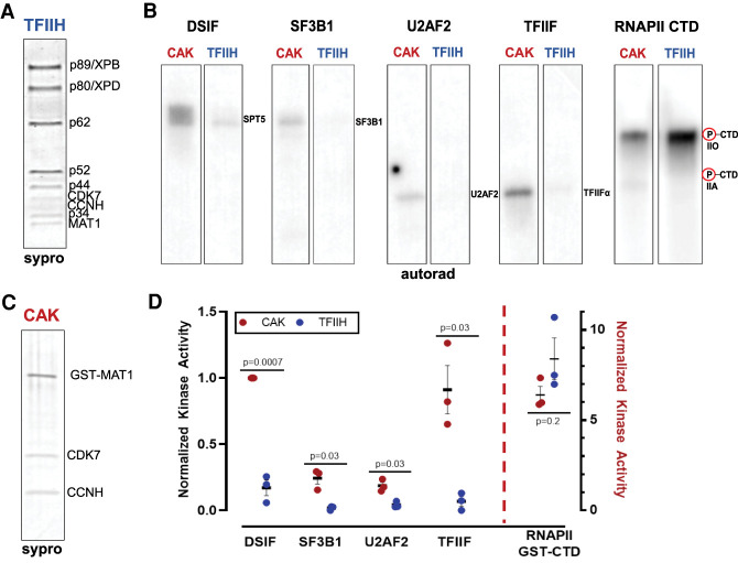
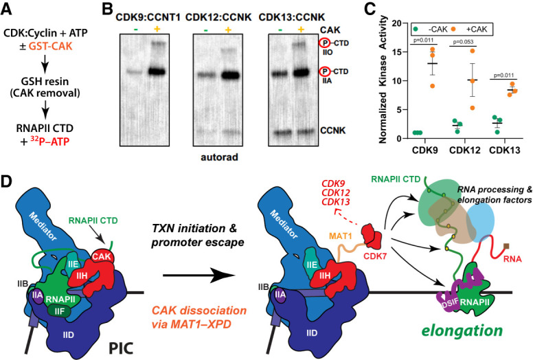
CDK7 associates with the 10-subunit TFIIH complex and regulates transcription by phosphorylating the C-terminal domain (CTD) of RNA polymerase II (RNAPII). Few additional CDK7 substrates are known. Here, using the covalent inhibitor SY-351 and quantitative phosphoproteomics, we identified CDK7 kinase substrates in human cells. Among hundreds of high-confidence targets, the vast majority are unique to CDK7 (i.e., distinct from other transcription-associated kinases), with a subset that suggest novel cellular functions. Transcription-associated factors were predominant CDK7 substrates, including SF3B1, U2AF2, and other splicing components. Accordingly, widespread and diverse splicing defects, such as alternative exon inclusion and intron retention, were characterized in CDK7-inhibited cells. Combined with biochemical assays, we establish that CDK7 directly activates other transcription-associated kinases CDK9, CDK12, and CDK13, invoking a "master regulator" role in transcription. We further demonstrate that TFIIH restricts CDK7 kinase function to the RNAPII CTD, whereas other substrates (e.g., SPT5 and SF3B1) are phosphorylated by the three-subunit CDK-activating kinase (CAK; CCNH, MAT1, and CDK7). These results suggest new models for CDK7 function in transcription and implicate CAK dissociation from TFIIH as essential for kinase activation. This straightforward regulatory strategy ensures CDK7 activation is spatially and temporally linked to transcription, and may apply toward other transcription-associated kinases.

Keywords: CDK12; CDK13; CDK7; CDK9; SF3B1; SILAC-MS; TFIIH; kinase inhibitor; splicing; transcription.


Figures

Figure 1.
Figure 1.
SY-351 is a potent and highly selective covalent inhibitor of human CDK7. (A) SY-351 structure. (B) Kinome selectivity in A549 cell lysate, with 0.2 µM and 1 µM SY-351. The top six hits shown are kinases inhibited > 50% by 1 µM SY-351. Note that SY-351 was used at 0.05 µM (50 nM) throughout this study. (C) CDK7 and CDK12 target occupancy in HL-60 cells after 1-h treatment. The SY-351 EC50 is 8.3 nM for CDK7 and 36 nM for CDK12. The SY-351 EC90 is 39 nM for CDK7 and 172 nM for CDK12. The dashed line indicates 50 nM SY-351, the dose used throughout this study. (D) SY-351 inhibition of active kinases CDK7/CCNH/MAT1, CDK2/CCNE1, CDK9/CCNT1, CDK12/CCNK at Km ATP for each enzyme. The best fit IC50 values are 23 nM, 321 nM, 226 nM, and 367 nM, respectively.
Figure 2.
Figure 2.
SILAC phosphoproteomic overview and identification of phosphorylation sites significantly changed by SY-351 treatment. (A) Experimental design and workflow using SILAC double-labeled HL-60 cells (Light:Heavy) and phosphoproteomics. Labeled cell populations were treated with vehicle (DMSO) or 50 nM SY-351 as indicated, with two biological replicates of SY-351-treated heavy cells (vs. DMSO light cells), a “label-flip” biological replicate of SY-351 light cells versus DMSO heavy cells, and a “null” condition for which both heavy and light were treated with DMSO. Tryptic peptides were enriched using titanium dioxide beads and analyzed by high-resolution mass spectrometry to identify phosphopeptides and quantify their light:heavy SILAC ratios. (B) Volcano plot showing the statistical significance and magnitude of change with SY-351 treatment for phosphorylation site SILAC ratios. The adjusted P-value range for phosphorylation site ratios is indicated in color. (C) Venn diagram showing phosphorylation sites shared or mutually exclusive for transcription-associated kinases CDK7, CDK8, CDK9, and CDK12/CDK13. For each kinase, the top 400 identified phosphorylated proteins, based upon the largest negative log fold change values and P < 0.05, were used for comparisons, and the number of overlapping proteins was calculated for all combinations of samples. CDK7 targets identified here were compared with proteins identified as substrates for CDK8 in HCT116 (Poss et al. 2016), CDK9 in Raji B lymphocytes (Decker et al. 2019), and CDK12/13 in IMR-32 and Kelly cells (Krajewska et al. 2019).
Figure 3.
Figure 3.
Protein–protein interaction network of phosphoproteins with significantly decreasing phosphorylation sites upon SY-351 treatment. Proteins with decreasing phosphorylation sites (log2FC < 0 and FDR < 0.05) were visualized using the STRING database web application, and clustered using the MCL algorithm, with clusters indicated by node color.
Figure 4.
Figure 4.
CDK7 inhibition with SY-351 causes widespread defects in splicing. (A) Differentially expressed mRNAs following SY351 treatment (DESeq2). Replicate RNA-seq data sets were generated from rRNA-depleted total RNA from HL60 cells treated with DMSO (CTRL) or SY-351 (50 nM in DMSO) for 5 h. (B) GSEA analysis. (C) SY-351 favors exon inclusion over exon skipping, splicing over retention of introns, and alters 5′ and 3′ splice site selection equally, without preference for upstream or downstream sites. Alternative splicing events significantly affected by SY-351 were identified in replicate RNA-seq data sets using MAJIQ (Vaquero-Garcia et al. 2016). (D) IGV genome browser Sashimi plot of a segment of the LTV1 gene. Normalized read numbers for DMSO control and SY-351 treated samples are shown on the Y-axis. Splice junction read numbers for sense strand transcripts are shown in blue and red. Note reduced exon inclusion (green arrow) and retention of both flanking introns (black arrows) in SY-351. (E) Immunofluorescence microscopy of endogenous SF3B1 (green) and Hoechst (blue) in DMSO and SY-351 conditions. (F) Quantitation of SF3B1 puncta per nucleus in DMSO and SY-351 conditions, P = 0.001 (n = 3 biological replicates).
Figure 5.
Figure 5.
CDK7 kinase activity is negatively regulated by TFIIH. (A) SYPRO-stained gel of human TFIIH. (B) TFIIH activates CDK7 kinase toward the CTD, but reduces or prevents CDK7 phosphorylation of DSIF, SF3B1, U2AF2, and TFIIF. Within the CAK, CDK7 is activated toward these substrates. (C) SYPRO-stained gel of human CAK complex (MAT1 is GST-tagged). (D) Quantitation of CDK7 kinase results across biological replicates (n = 3) with autorad signal normalized to CAK phosphorylated DSIF; P-values as shown.
Figure 6.
Figure 6.
CDK7 is a master regulator of transcription-associated kinases; model. (A) Experimental overview; CDK:cyclin complexes were incubated with the CAK, followed by CAK removal. CDK:cyclin complexes were then tested for activity against the RNAPII CTD. (B) Representative kinase data in which purified CDK9:CCNT1, CDK12:CCNK, or CDK13:CCNK were tested against a common substrate, the RNAPII CTD. Each kinase complex was prephosphorylated by CDK7 (vs. controls) as shown. (C) Quantitation of kinase results across biological replicates (n = 3) with autorad signal normalized to CDK9 CAK pretreatment; P-values as shown. (D) Working model for TFIIH/CDK7 function in RNAPII transcription. Within the PIC, the CAK is assembled with TFIIH and CDK7 is activated toward the RNAPII CTD. The CDK7 phosphorylated CTD can be released from Mediator (Søgaard and Svejstrup 2007) and helps recruit chromatin modifiers, capping enzymes, and other RNA-processing factors (Ebmeier et al. 2017). Following promoter escape, the CAK may dissociate from TFIIH while maintaining interaction with XPD in the TFIIH core, via the flexible CAK subunit MAT1 (Greber et al. 2019). This “CAK release” allows access to cotranscriptional substrates (e.g., splicing and elongation factors) and also activates CDK7 toward these substrates. In this way, CDK7 activity is spatially and temporally regulated by TFIIH. Moreover, CDK7-dependent activation of CDK9, CDK12, and CDK13 would promote coordinated regulation of promoter-proximal pausing, transcription elongation, and mRNA biogenesis.

References

    1. Anders S, Reyes A, Huber W. 2012. Detecting differential usage of exons from RNA-seq data. Genome Res 22: 2008–2017. 10.1101/gr.133744.111 - DOI - PMC - PubMed
    1. Bartkowiak B, Greenleaf AL. 2015. Expression, purification, and identification of associated proteins of the full-length hCDK12/CyclinK complex. J Biol Chem 290: 1786–1795. 10.1074/jbc.M114.612226 - DOI - PMC - PubMed
    1. Begley MJ, Yun CH, Gewinner CA, Asara JM, Johnson JL, Coyle AJ, Eck MJ, Apostolou I, Cantley LC. 2015. EGF-receptor specificity for phosphotyrosine-primed substrates provides signal integration with Src. Nat Struct Mol Biol 22: 983–990. 10.1038/nsmb.3117 - DOI - PMC - PubMed
    1. Bentley DL. 2014. Coupling mRNA processing with transcription in time and space. Nature reviews Genetics 15: 163–175. 10.1038/nrg3662 - DOI - PMC - PubMed
    1. Bösken CA, Farnung L, Hintermair C, Merzel Schachter M, Vogel-Bachmayr K, Blazek D, Anand K, Fisher RP, Eick D, Geyer M. 2014. The structure and substrate specificity of human Cdk12/Cyclin K. Nat Commun 5: 3505 10.1038/ncomms4505 - DOI - PMC - PubMed

Publication types

MeSH terms

Substances

LinkOut - more resources