. 2020 Oct;8(2):e000940.
doi: 10.1136/jitc-2020-000940.

In situ immunization of a TLR9 agonist virus-like particle enhances anti-PD1 therapy

Affiliations

In situ immunization of a TLR9 agonist virus-like particle enhances anti-PD1 therapy

Yinwen Cheng et al. J Immunother Cancer. 2020 Oct.

Abstract

Background: CMP-001 is a novel Toll-like receptor-9 agonist that consists of an unmethylated CpG-A motif-rich G10 oligodeoxynucleotide (ODN) encapsulated in virus-like particles. In situ vaccination of CMP-001 is believed to activate local tumor-associated plasmacytoid dendritic cells (pDCs) leading to type I interferon secretion and tumor antigen presentation to T cells and systemic antitumor T cell responses. This study is designed to investigate if CMP-001 would enhance head and neck squamous cell carcinoma (HNSCC) tumor response to anti-programmed cell death protein-1 (anti-PD-1) therapy in a human papilloma virus-positive (HPV+) tumor mouse model.

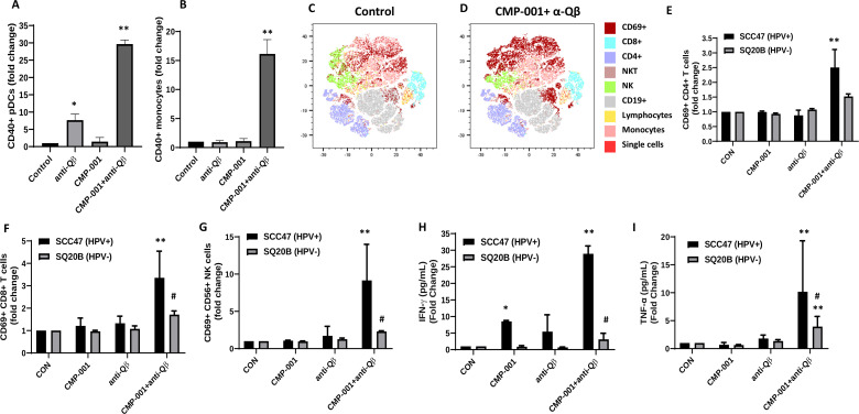
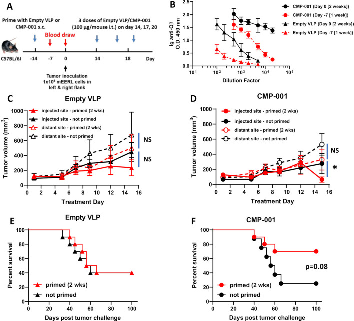
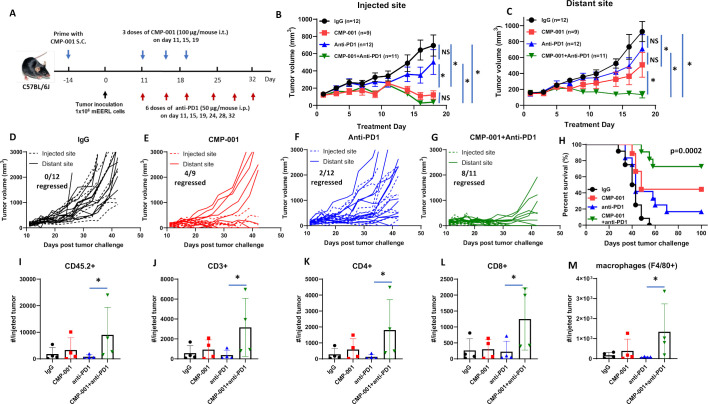
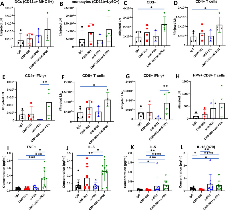
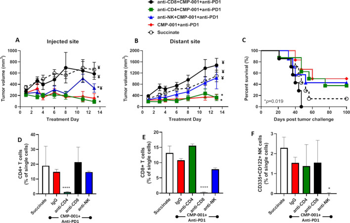
Methods: Immune cell activation in response to CMP-001±anti-Qβ was performed using co-cultures of peripheral blood mononuclear cells and HPV+/HPV- HNSCC cells and then analyzed by flow cytometry. In situ vaccination with CMP-001 alone and in combination with anti-PD-1 was investigated in C57BL/6 mice-bearing mEERL HNSCC tumors and analyzed for anti-Qβ development, antitumor response, survival and immune cell recruitment. The role of antitumor immune response due to CMP-001+anti-PD-1 treatment was investigated by the depletion of natural killer (NK), CD4+ T, and CD8+ T cells.

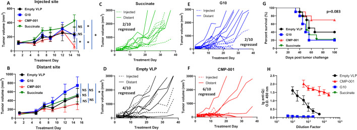
Results: Results showed that the activity of CMP-001 on immune cell (pDCs, monocytes, CD4+/CD8+ T cells and NK cells) activation depends on the presence of anti-Qβ. A 2-week 'priming' period after subcutaneous administration of CMP-001 was required for robust anti-Qβ development in mice. In situ vaccination of CMP-001 was superior to unencapsulated G10 CpG-A ODN at suppressing both injected and uninjected (distant) tumors. In situ vaccination of CMP-001 in combination with anti-PD-1 therapy induced durable tumor regression at injected and distant tumors and significantly prolonged mouse survival compared with anti-PD-1 therapy alone. The antitumor effect of CMP-001+anti-PD-1 was accompanied by increased interferon gamma (IFNγ)+ CD4+/CD8+ T cells compared with control-treated mice. The therapeutic and abscopal effect of CMP-001+ anti-PD-1 therapy was completely abrogated by CD8+ T cell depletion.

Conclusions: These results demonstrate that in situ vaccination with CMP-001 can induce both local and abscopal antitumor immune responses. Additionally, the antitumor efficacy of CMP-001 combined with α-PD-1 therapy warrants further study as a novel immunotherapeutic strategy for the treatment of HNSCC.

Keywords: adjuvants; dendritic cells; head and neck neoplasms; immunologic; immunotherapy; vaccination.


Conflict of interest statement

Competing interests: None declared.

Figures

Figure 1
Figure 1
Activity of CMP-001 on immune cell infiltration depends on the presence of anti-Qβ. (A, B) Peripheral blood mononuclear cells (PBMCs) were treated with CMP-001 (5 µg/mL) and/or anti-Qβ (5 µg/mL) for 24 hours. Activation of plasmacytoid dendritic cells (pDCs) (CD45+ CD3- CD19- CD11c- BDCA-4+) (A) and monocytes (CD45+ CD3- CD19- CD11c+ CD14+) (B) was monitored by flow cytometry. (C–I) SQ20B (human papilloma virus-negative (HPV-)) and SCC47 (human papilloma virus-positive (HPV+)) cells were co-cultured with PBMCs, treated with CMP-001±anti-Qβ as previously described, then flow cytometry and T-distributed Stochastic Neighbor Embedding (t-SNE) data analysis (C, D) was used to identify activated CD4+ (E), CD8+ (F), and natural killer (NK) cells (G), and interferon gamma (IFNγ) (H) and tumor necrosis factor α (TNFα) (I) concentrations in cell culture media analyzed by ELISA. Succinate buffer was used as a control (CON). Representative t-SNE plots are shown for CON (C) and CMP-001+anti-Qβ (D). Bar graphs shown represent the mean of n=3 experiments. Error bars represent SD from the mean. *p<0.05 versus CON; **p<0.05 versus CMP-001; #p<0.05 versus UM-SCC47.
Figure 2
Figure 2
A 2-week ‘priming’ is required for robust anti-Qβ development and favorable survival outcomes. (A) The treatment schema for the priming of C57BL/6 mice (n=10/treatment group) with empty virus-like particle (VLP) or CMP-001 (100 µg/mouse) subcutaneously (s.c.) 2 weeks or 1 week before tumor challenge with mEERL cells on both left and right flanks. (B) Blood samples from a subset of mice (n=4–5) were collected 1 week (Day −7) and 2 weeks (Day 0) after priming and analyzed for immunoglobulin (Ig) anti-Qβ concentration. After tumor formation, empty VLP or CMP-001 was then administered to the left tumor only intratumorally (i.t.) every 3 days. Tumor volume (C, D) for the left (injected) and right (distant (uninjected)) tumors in primed and unprimed empty VLP (C) and CMP-001 (D) treated mice was measured three to four times per week. Tumor growth curves shown were terminated when a mouse in any treatment group reached euthanasia criteria. Overall survival was analyzed by constructing Kaplan-Meier plots for empty VLP (E) and CMP-001 (F) treated mice. Error bars represent SD from the mean. *p<0.05. NS, non-significant. Note: Treatment Day 1 in tumor growth graphs (C, D) represent Day 14 in treatment schema in (A).
Figure 3
Figure 3
CMP-001 showed superior antitumor effect in vivo compared with G10. mEERL tumor-bearing C57BL/6 mice (n=10/treatment group) were treated intratumorally with succinate as a control, G10 CpG ODN, empty virus-like particle (VLP) (100 µg/mouse) and CMP-001 (100 µg/mouse) into the left tumor on Days 14, 17 and 20 after mEERL tumor inoculation. All mice were primed 2 weeks before tumor inoculation with their respective assigned treatments. Tumor volume for the left (injected) (A) and right (distant (uninjected)) (B) tumors in the treated mice was measured three to four times per week. Tumor growth curves shown were terminated when a mouse in any treatment group reached euthanasia criteria. Spaghetti plots of all mice in each of the treatment groups are shown in (C)–(F). Numbers indicated in the spaghetti plots are the number of distant tumors that regressed. Overall survival was analyzed by constructing Kaplan-Meier plots (G) for the treated mice. Mouse serum from a subset of mice (n=4–5 mice/treatment group) was collected 2 weeks after priming for analysis of immunoglobulin (Ig) anti-Qβ concentration (H). Error bars represent SD from the mean. *p<0.05. NS, non-significant.
Figure 4
Figure 4
CMP-001 enhances anti-programmed cell death protein-1 (anti-PD1) therapy. (A) mEERL tumor-bearing C57Bl/6 mice (n=9–12/treatment group) were treated with immunoglobulin G (IgG) (+succinate) as a control, CMP-001 (+IgG), anti-PD1 (+succinate) and CMP-001+anti-PD1. CMP-001 and succinate were administered intratumorally into the left tumor on Days 11, 15, and 19, and anti-PD1 was administered intraperitoneally on Days 11, 15, 19, 24, 28, and 32 after mEERL tumor inoculation. All mice receiving CMP-001 were primed 2 weeks before tumor innoculation. Tumor volume for the left (injected) (B) and right (distant (uninjected)) (C) tumors in the treated mice was measured three to four times per week. Tumor growth curves shown were terminated when a mouse in any treatment group reached euthanasia criteria. Spaghetti plots of all mice in each of the treatment groups are shown in (D)–(G) Numbers indicated in the spaghetti plots are the number of distant tumors that regressed. Overall survival was analyzed by constructing Kaplan-Meier plots (H) for the treated mice. A subset of mice (n=3–4 mice/group) from each treatment were euthanized 2 days after their last treatment, and tumors at the injected tumor site were harvested and analyzed by flow cytometry for select immune subsets such as CD45.2+ cells (I), CD3+ (J), CD4+ (K), and CD8+ (L) T cells and macrophages (M). Error bars represent SD from the mean. *p<0.05. NS, non-significant.
Figure 5
Figure 5
CMP-001 combined with anti-programmed cell death protein-1 (anti-PD1) increases immune cell infiltration to the draining lymph node (LN). A subset of mice (n=3–4 mice/group) from each treatment group from the experiment shown in figure 4A–C were euthanized 2 days after their last drug treatment and lingual LNs at the injected tumor site were harvested and analyzed by flow cytometry for select immune subsets such as dendritic cells (DCs) (A), monocytes (B), CD3+ (C), CD4+ (D), IFNγ+ CD4+ (E), CD8+ (F), IFNγ+ CD8+ (G) and HPV+ CD8+ (H) T cells. Serum samples were collected for cytokine analysis using a murine cytokine multiplex assay. Shown are concentration (µg/mL) levels for TNFα (I), IL-6 (J), IL-5 (K) and IL-12(p70) (L). Error bars represent SD from the mean. *p<0.05; **p<0.01; ***p<0.001; ****p<0.0001. HPV+, human papilloma virus-positive; IFNγ, interferon gamma; IgG, immunoglobulin G; IL, interleukin.
Figure 6
Figure 6
The antitumor effects of CMP-001+α-PD1 depends on CD8+ T cells. mEERL tumor-bearing C57Bl/6 mice (n=10–12/treatment group) were treated with CMP-001+anti-programmed cell death protein-1 (anti-PD1) as described in figure 4 with or without anti-CD4, anti-CD8 or anti–asialo-GM1 (anti-NK) antibodies 1 and 3 days prior to tumor inoculation, and every 3–4 days after tumor inoculation. Succinate buffer was used as non-treatment control. All mice receiving CMP-001 were primed 2 weeks before tumor inoculation. Tumor volume for the left (injected) (A) and right (distant (uninjected)) (B) tumors in the treated mice was measured three to four times per week. Tumor growth curves shown were terminated when a mouse in any treatment group reached euthanasia criteria. Overall survival was analyzed by constructing Kaplan-Meier plots (C) for the treated mice. *p<0.05 versus succinate, ¥p<0.05 versus CMP-001+ anti-PD1. (D–F) Splenic peripheral blood mononuclear cells (PBMCs) were isolated from a subset of mice (n=3) 1 day after the last treatment and were analyzed by flow cytometry for validation of CD4+ T cell (D), CD8+ T cell (E), and natural (NK) cell (F) depletion. *p<0.05; ****p<0.0001. Error bars represent SD from the mean. IgG, immunoglobulin G; NK, natural killer.
Figure 7
Figure 7
TLR9 gene expression positively correlates with overall survival of patients with head and neck squamous cell carcinoma (HNSCC) and T cell infiltration. (A) Kaplan-Meier survival curves comparing overall survival of patients with HNSCC (n=520) created from The Cancer Genome Atlas (TCGA) database according to high (n=284) and low (n=236) TLR9 tumor gene expression. (B, C) Dot plots illustrate the percentage of CD8+ (B) and activated memory CD4+ (C) tumor-infiltrating T cells in HNSCC patients with high and low TLR9 expression from (A). Error bars represent SD from the mean. **p<0.01; ****p<0.0001.

Similar articles

Cited by

References

    1. Bauml J, Seiwert TY, Pfister DG, et al. . Pembrolizumab for platinum- and Cetuximab-Refractory head and neck cancer: results from a single-arm, phase II study. J Clin Oncol 2017;35:1542–9. 10.1200/JCO.2016.70.1524 - DOI - PMC - PubMed
    1. Burtness B, Harrington KJ, Greil R, et al. . KEYNOTE-048: phase III study of first-line pembrolizumab (P) for recurrent/metastatic head and neck squamous cell carcinoma (r/m HNSCC). Ann Oncol 2018;29:viii729 10.1093/annonc/mdy424.045 - DOI
    1. Mehra R, Seiwert TY, Gupta S, et al. . Efficacy and safety of pembrolizumab in recurrent/metastatic head and neck squamous cell carcinoma: pooled analyses after long-term follow-up in KEYNOTE-012. Br J Cancer 2018;119:153–9. 10.1038/s41416-018-0131-9 - DOI - PMC - PubMed
    1. Fares CM, Van Allen EM, Drake CG, et al. . Mechanisms of resistance to immune checkpoint blockade: why does checkpoint inhibitor immunotherapy not work for all patients? Am Soc Clin Oncol Educ Book 2019;39:147–64. 10.1200/EDBK_240837 - DOI - PubMed
    1. Steinman RM. The dendritic cell system and its role in immunogenicity. Annu Rev Immunol 1991;9:271–96. 10.1146/annurev.iy.09.040191.001415 - DOI - PubMed

Publication types

MeSH terms