

Solitary pulmonary nodule imaging approaches and the role of optical fibre-based technologies
- PMID: 33060152
- PMCID: PMC8174723
- DOI: 10.1183/13993003.02537-2020
Solitary pulmonary nodule imaging approaches and the role of optical fibre-based technologies
Abstract
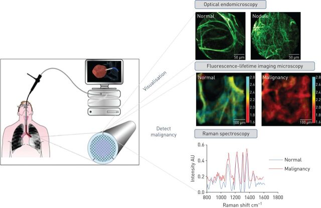
Solitary pulmonary nodules (SPNs) are a clinical challenge, given there is no single clinical sign or radiological feature that definitively identifies a benign from a malignant SPN. The early detection of lung cancer has a huge impact on survival outcome. Consequently, there is great interest in the prompt diagnosis, and treatment of malignant SPNs. Current diagnostic pathways involve endobronchial/transthoracic tissue biopsies or radiological surveillance, which can be associated with suboptimal diagnostic yield, healthcare costs and patient anxiety. Cutting-edge technologies are needed to disrupt and improve, existing care pathways. Optical fibre-based techniques, which can be delivered via the working channel of a bronchoscope or via transthoracic needle, may deliver advanced diagnostic capabilities in patients with SPNs. Optical endomicroscopy, an autofluorescence-based imaging technique, demonstrates abnormal alveolar structure in SPNs in vivo Alternative optical fingerprinting approaches, such as time-resolved fluorescence spectroscopy and fluorescence-lifetime imaging microscopy, have shown promise in discriminating lung cancer from surrounding healthy tissue. Whilst fibre-based Raman spectroscopy has enabled real-time characterisation of SPNs in vivo Fibre-based technologies have the potential to enable in situ characterisation and real-time microscopic imaging of SPNs, which could aid immediate treatment decisions in patients with SPNs. This review discusses advances in current imaging modalities for evaluating SPNs, including computed tomography (CT) and positron emission tomography-CT. It explores the emergence of optical fibre-based technologies, and discusses their potential role in patients with SPNs and suspected lung cancer.
Copyright ©ERS 2021.
Conflict of interest statement
Conflict of interest: S. Fernandes reports grants from MRC and Boston Scientific, during the conduct of the study. Conflict of interest: G. Williams has patents planned relating to FLIM. Conflict of interest: E. Williams has patents planned relating to FLIM. Conflict of interest: K. Ehrlich has nothing to disclose. Conflict of interest: J. Stone reports grants from EPSRC and Boston Scientific, during the conduct of the study; and has a patent imaging fibre issued. Conflict of interest: N. Finlayson reports grants from EPSRC, during the conduct of the study; other from Prothea-X, outside the submitted work. Conflict of interest: M. Bradley reports grants from MRC, Boston Scientific and EPSRC, during the conduct of the study. Conflict of interest: R.R. Thomson reports grants from University of Edinburgh, during the conduct of the study. Conflict of interest: A.R. Akram reports academic grants, during the conduct of the study. Conflict of interest: K. Dhaliwal reports grants from MRC, Boston Scientific and EPSRC, and reimbursment for travel and consultancy from Mauna Kea Technologies, during the conduct of the study.
Figures
References
Publication types
MeSH terms
Grants and funding
LinkOut - more resources
Full Text Sources
Medical