

Interrogation of kinase genetic interactions provides a global view of PAK1-mediated signal transduction pathways
- PMID: 33060198
- PMCID: PMC7863907
- DOI: 10.1074/jbc.RA120.014831
Interrogation of kinase genetic interactions provides a global view of PAK1-mediated signal transduction pathways
Abstract
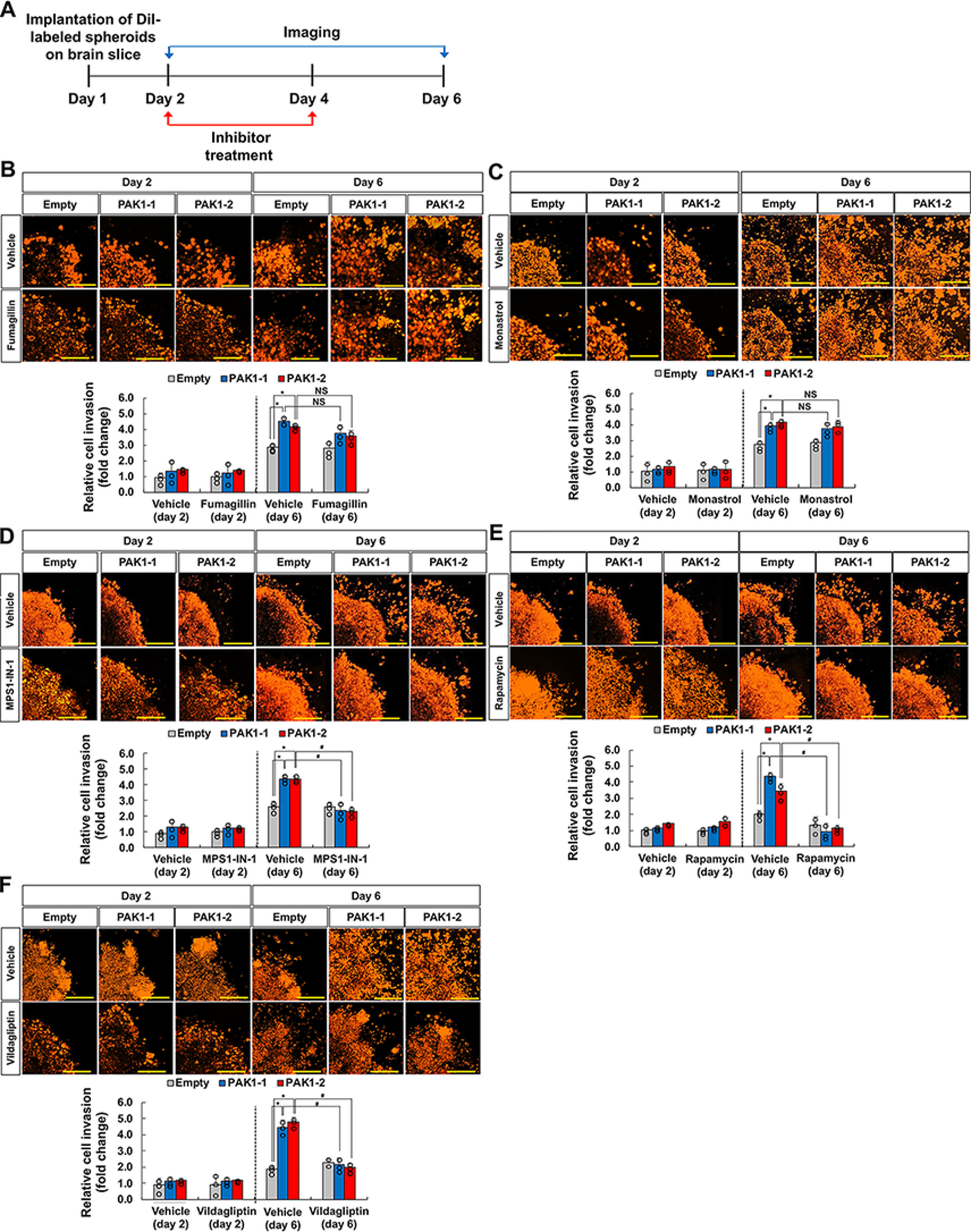
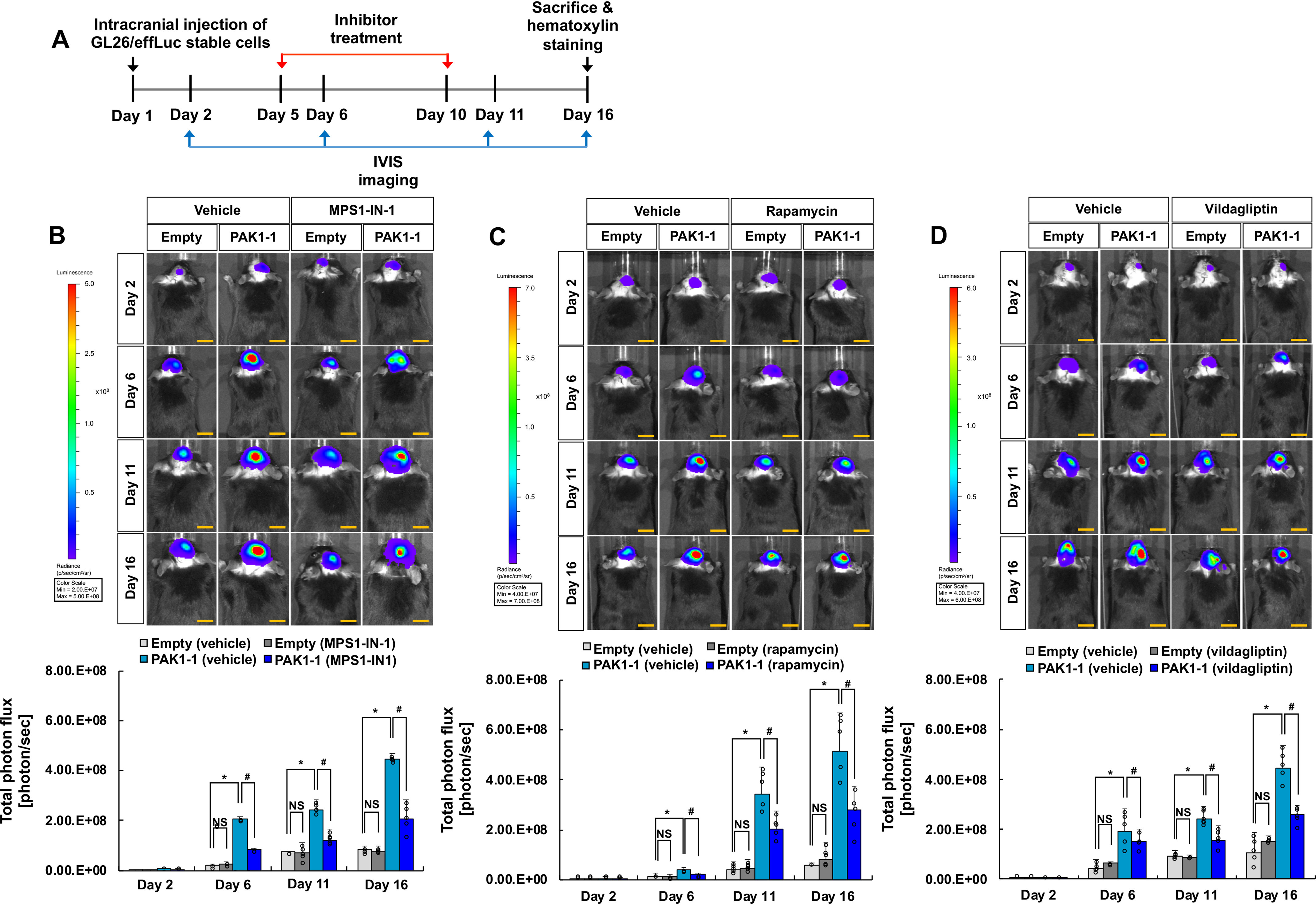
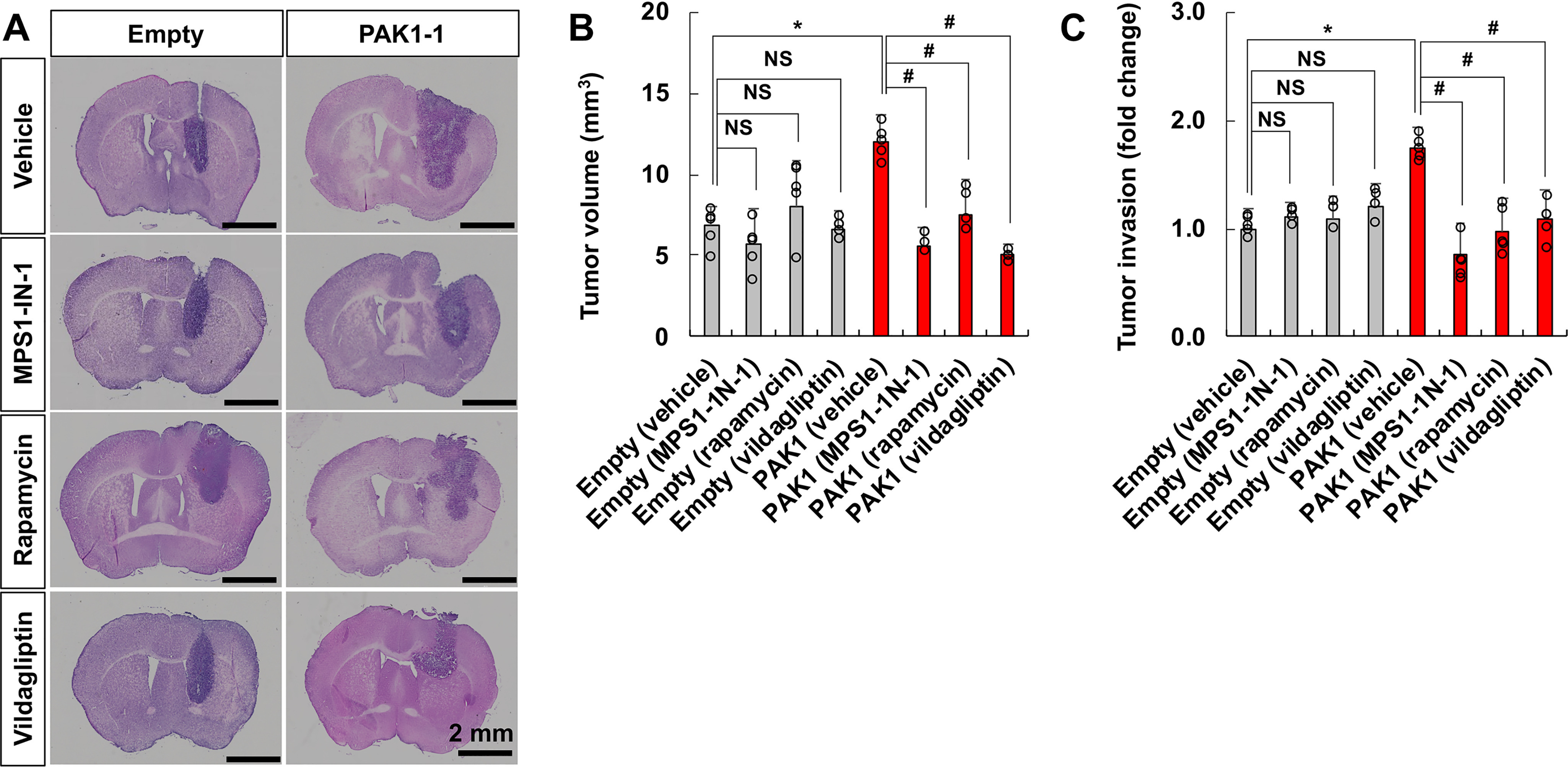
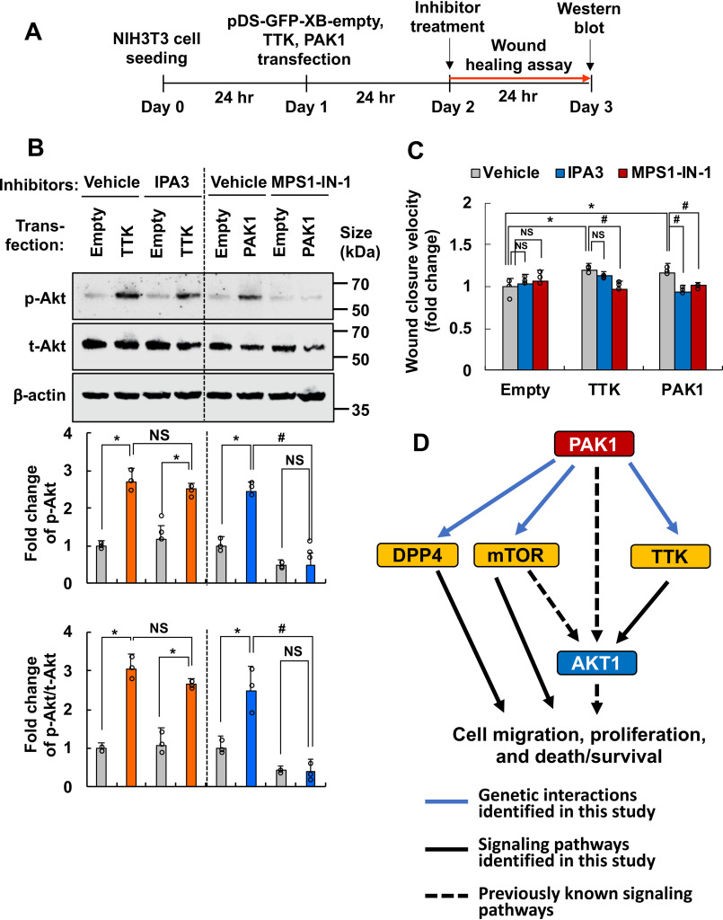
Kinases are critical components of intracellular signaling pathways and have been extensively investigated with regard to their roles in cancer. p21-activated kinase-1 (PAK1) is a serine/threonine kinase that has been previously implicated in numerous biological processes, such as cell migration, cell cycle progression, cell motility, invasion, and angiogenesis, in glioma and other cancers. However, the signaling network linked to PAK1 is not fully defined. We previously reported a large-scale yeast genetic interaction screen using toxicity as a readout to identify candidate PAK1 genetic interactions. En masse transformation of the PAK1 gene into 4,653 homozygous diploid Saccharomyces cerevisiae yeast deletion mutants identified ∼400 candidates that suppressed yeast toxicity. Here we selected 19 candidate PAK1 genetic interactions that had human orthologs and were expressed in glioma for further examination in mammalian cells, brain slice cultures, and orthotopic glioma models. RNAi and pharmacological inhibition of potential PAK1 interactors confirmed that DPP4, KIF11, mTOR, PKM2, SGPP1, TTK, and YWHAE regulate PAK1-induced cell migration and revealed the importance of genes related to the mitotic spindle, proteolysis, autophagy, and metabolism in PAK1-mediated glioma cell migration, drug resistance, and proliferation. AKT1 was further identified as a downstream mediator of the PAK1-TTK genetic interaction. Taken together, these data provide a global view of PAK1-mediated signal transduction pathways and point to potential new drug targets for glioma therapy.
Keywords: PAK1; cell migration; cell proliferation; drug resistance; genetic interaction; glioma; kinase; molecular cell biology; signal transduction.
© 2020 Kim et al.
Conflict of interest statement
Conflict of interest—The authors declare that they have no conflicts of interest with the contents of this article.
Figures
Similar articles
-
PAK1 Inhibition Suppresses the Proliferation, Migration and Invasion of Glioma Cells.Curr Protein Pept Sci. 2023;24(2):178-189. doi: 10.2174/1389203724666221226150329. Curr Protein Pept Sci. 2023. PMID: 36573046
-
P21-activated kinase 1 stimulates colon cancer cell growth and migration/invasion via ERK- and AKT-dependent pathways.Biochim Biophys Acta. 2010 Sep;1803(9):1106-13. doi: 10.1016/j.bbamcr.2010.05.007. Epub 2010 Jun 1. Biochim Biophys Acta. 2010. PMID: 20595063
-
p21-activated kinase 1 determines stem-like phenotype and sunitinib resistance via NF-κB/IL-6 activation in renal cell carcinoma.Cell Death Dis. 2015 Feb 12;6(2):e1637. doi: 10.1038/cddis.2015.2. Cell Death Dis. 2015. PMID: 25675297 Free PMC article.
-
Current trends and opportunities in targeting p21 activated kinase-1(PAK1) for therapeutic management of breast cancers.Gene. 2020 Nov 15;760:144991. doi: 10.1016/j.gene.2020.144991. Epub 2020 Jul 25. Gene. 2020. PMID: 32717309 Review.
-
P21-Activated Kinase 1: Emerging biological functions and potential therapeutic targets in Cancer.Theranostics. 2020 Aug 1;10(21):9741-9766. doi: 10.7150/thno.46913. eCollection 2020. Theranostics. 2020. PMID: 32863957 Free PMC article. Review.
Cited by
-
PAK4-relevant proliferation reduced by cell autophagy via p53/mTOR/p-AKT signaling.Transl Cancer Res. 2023 Mar 31;12(3):461-472. doi: 10.21037/tcr-22-2272. Epub 2023 Mar 21. Transl Cancer Res. 2023. PMID: 37033362 Free PMC article.
-
Regulation of Vascular Injury and Repair by P21-Activated Kinase 1 and P21-Activated Kinase 2: Therapeutic Potential and Challenges.Biomolecules. 2024 Dec 13;14(12):1596. doi: 10.3390/biom14121596. Biomolecules. 2024. PMID: 39766303 Free PMC article. Review.
-
Identification of Genetic Modifiers of TDP-43: Inflammatory Activation of Astrocytes for Neuroinflammation.Cells. 2021 Mar 18;10(3):676. doi: 10.3390/cells10030676. Cells. 2021. PMID: 33803845 Free PMC article.
References
-
- Teramoto, H., Crespo, P., Coso, O. A., Igishi, T., Xu, N., and Gutkind, J. S. (1996) The small GTP-binding protein Rho activates c-Jun N-terminal kinases/stress-activated protein kinases in human kidney 293T cells: evidence for a Pak-independent signaling pathway. J. Biol. Chem. 271, 25731–25734 10.1074/jbc.271.42.25731 - DOI - PubMed
Publication types
MeSH terms
Substances
Grants and funding
LinkOut - more resources
Full Text Sources
Molecular Biology Databases
Research Materials
Miscellaneous
