Review
. 2020 Oct 16;370(6514):eabb2153.
doi: 10.1126/science.abb2153.

Transient cortical circuits match spontaneous and sensory-driven activity during development

Affiliations
Review

Transient cortical circuits match spontaneous and sensory-driven activity during development

Zoltán Molnár et al. Science. .

Abstract

At the earliest developmental stages, spontaneous activity synchronizes local and large-scale cortical networks. These networks form the functional template for the establishment of global thalamocortical networks and cortical architecture. The earliest connections are established autonomously. However, activity from the sensory periphery reshapes these circuits as soon as afferents reach the cortex. The early-generated, largely transient neurons of the subplate play a key role in integrating spontaneous and sensory-driven activity. Early pathological conditions-such as hypoxia, inflammation, or exposure to pharmacological compounds-alter spontaneous activity patterns, which subsequently induce disturbances in cortical network activity. This cortical dysfunction may lead to local and global miswiring and, at later stages, can be associated with neurological and psychiatric conditions.


Conflict of interest statement

Competing interests: Authors declare no competing interests.

Figures

Fig. 1.
Fig. 1.. Cellular interactions in developing brain.
A: Cross-section of 18 gestational weeks human brain (2) (blue: subplate SP; pink: germinal zones (ventricular zone, VZ; subventricular zone, SVZ)). B: Schematic illustration of cellular cortical components present. Dividing radial glial progenitors in VZ are in contact with cerebrospinal fluid and receive endocrine signals, some through blood vessels (C). Immature neurons interact via paracrine (D) and autocrine (E) mechanisms, or couple into local networks via electrical (G) and chemical (H) synapses (F). These various forms of communications co-exist in the developing brain.
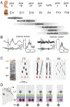
Fig. 2.
Fig. 2.. Corticogenesis and relationship with spontaneous activity patterns.
(A) Approximate time points of major developmental events in human neocortex at postconceptional weeks (pcw) and postnatal months (pnm) and in mouse cortex at embryonic (E) and postnatal (P) days (12). (B) Delta brush EEG activity and average Fast-Fourier transformation (FFT) spectrum evoked by hand movement (vertical bar) (13) in preterm human or following whisker stimulation in newborn rodent (vertical bar). Note similarity in spectrum. (C) Schematic illustration of developmental changes in spontaneous activity. (1) CR and subplate neurons (yellow and red bars, respectively) already discharge faster action potentials and at higher frequency than CP neurons (black bars). (2) Neurons electrically-coupled via gap junctions either generate local synchronized activity or propagating activity waves. (3) Discharges become faster and local networks discharge in synchronized bursts. Transient early-born neurons start to disappear during this phase. (4) Appearance of adult-like sparse desynchronized activity independent of transient neurons and circuits. (D) Subplate neurons are gap-junction-coupled when thalamocortical projections arrive. Thalamic fibers first establish synapses with subplate neurons before innervating L4 neurons. Subplate and L4 connections transiently co-exist to reinforce the more permanent thalamic projections that remain after subplate neurons lose their contact with thalamic projections, and also lose their contact to L4 themselves. Few L6b neurons survive to adulthood (14).
Fig. 3.
Fig. 3.. Transient circuit topologies during thalamocortical development.
A. Evoked responses in subplate and L4 after stimulation of the optic radiation in cat (45), of thalamus in mouse thalamocortical slices (15), and in vivo in ferret (39). Responses emerge and latencies are always shortest in SP. B. Topography is emerging in the subplate. Plotted is the difference in correlation of tuning curves between neighboring recording sites. Early-evoked responses in subplate show differencing responses at larger distances (39). C: Left: The integration of subplate neurites shows an age-specific pattern (57). Middle: Subplate axons target the septa in S1 barrel cortex (52). Right: Ablating a row of whiskers at birth changed the distribution of the corresponding neurites (57). D: Various transient connections only present during specific stages of development and not in the adult (34, 40, 59).
Fig. 4.
Fig. 4.. Establishment and plasticity of thalamo-cortical-thalamic circuits.
A. Development of thalamocortical connectivity in mouse (E15, P4 and P56). Thalamocortical projections cross the pallial-subpallial boundary simultaneously with corticothalamic projections, co-fasciculate, providing mutual guidance, and accumulate in subplate or outside the thalamus, possibly in TRN. L5 projections give side-branches selectively to higher order thalamic nuclei. L6 projections innervate both nuclei. B. Selective innervation of first and higher order thalamic nuclei. White circles illustrate dLGN and VB. L5 and L6b projections selectively innervate higher order thalamic nuclei, whereas L6a lack such preference (51, 63, 66). L5 terminals from S1 to PO are larger than L6a or L6b terminals from S1 to VB (66). C. Reciprocal thalamocortical connectivity during development and in adult. D. After sensory loss, cortical and thalamic connectivity in primary (1) and secondary (2) cortical area is changed.
Fig. 5.
Fig. 5.. Spontaneous activity in preterm and adult schizophrenics.
A, resting state fMRI (92). B, resting state EEG connectivity matrix at 8–15 Hz, showing stronger connectivity (red and yellow) in preterm (93). C, resting state fMRI data showing significant differences in thalamic connectivity between healthy (CON) and individuals with schizophrenia (SCZ) (94).

References

    1. Molnar Z et al., New insights into the development of the human cerebral cortex. J Anat 235, 432–451 (2019). - PMC - PubMed
    1. Adorjan I et al., Neuroserpin expression during human brain development and in adult brain revealed by immunohistochemistry and single cell RNA sequencing. J Anat 235, 543–554 (2019). - PMC - PubMed
    1. Katz LC, Shatz CJ, Synaptic activity and the construction of cortical circuits. Science 274, 1133–1138 (1996). - PubMed
    1. Wong FK et al., Pyramidal cell regulation of interneuron survival sculpts cortical networks. Nature 557, 668–673 (2018). - PMC - PubMed
    1. Riva M et al., Activity-dependent death of transient Cajal-Retzius neurons is required for functional cortical wiring. Elife 8, (2019). - PMC - PubMed

Publication types