. 2020 Oct 16;370(6514):327-331.
doi: 10.1126/science.abc0033.

Engineering synthetic morphogen systems that can program multicellular patterning

Affiliations

Engineering synthetic morphogen systems that can program multicellular patterning

Satoshi Toda et al. Science. .

Abstract

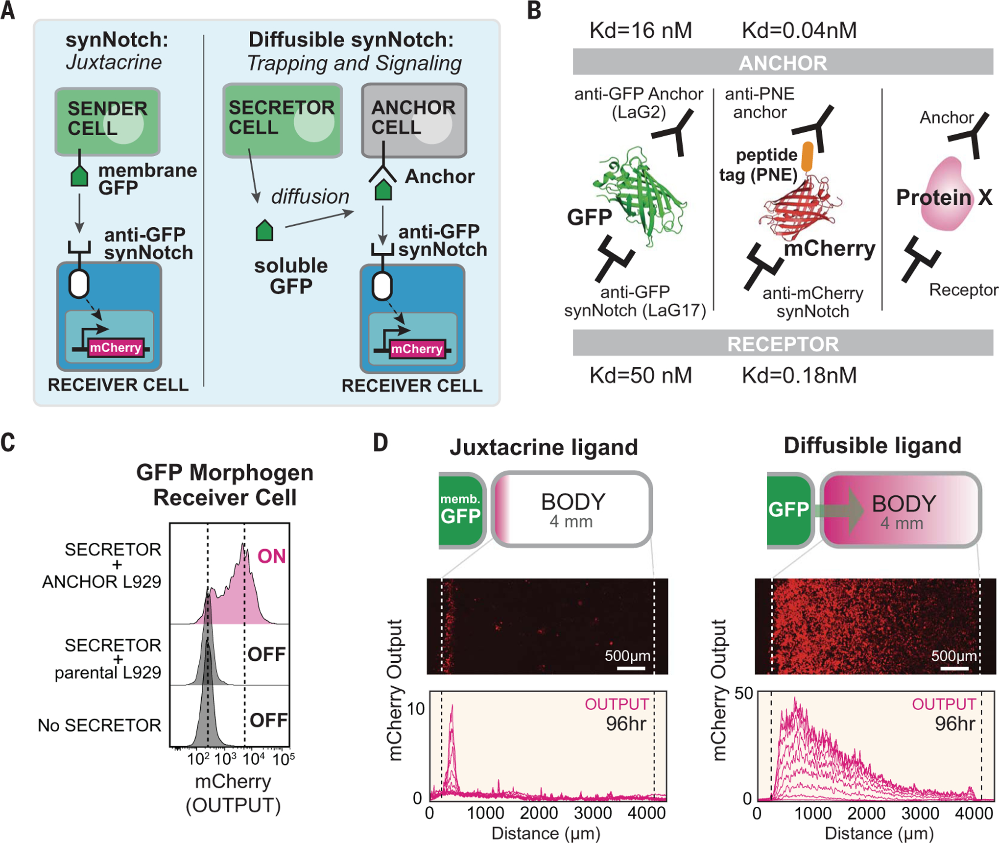
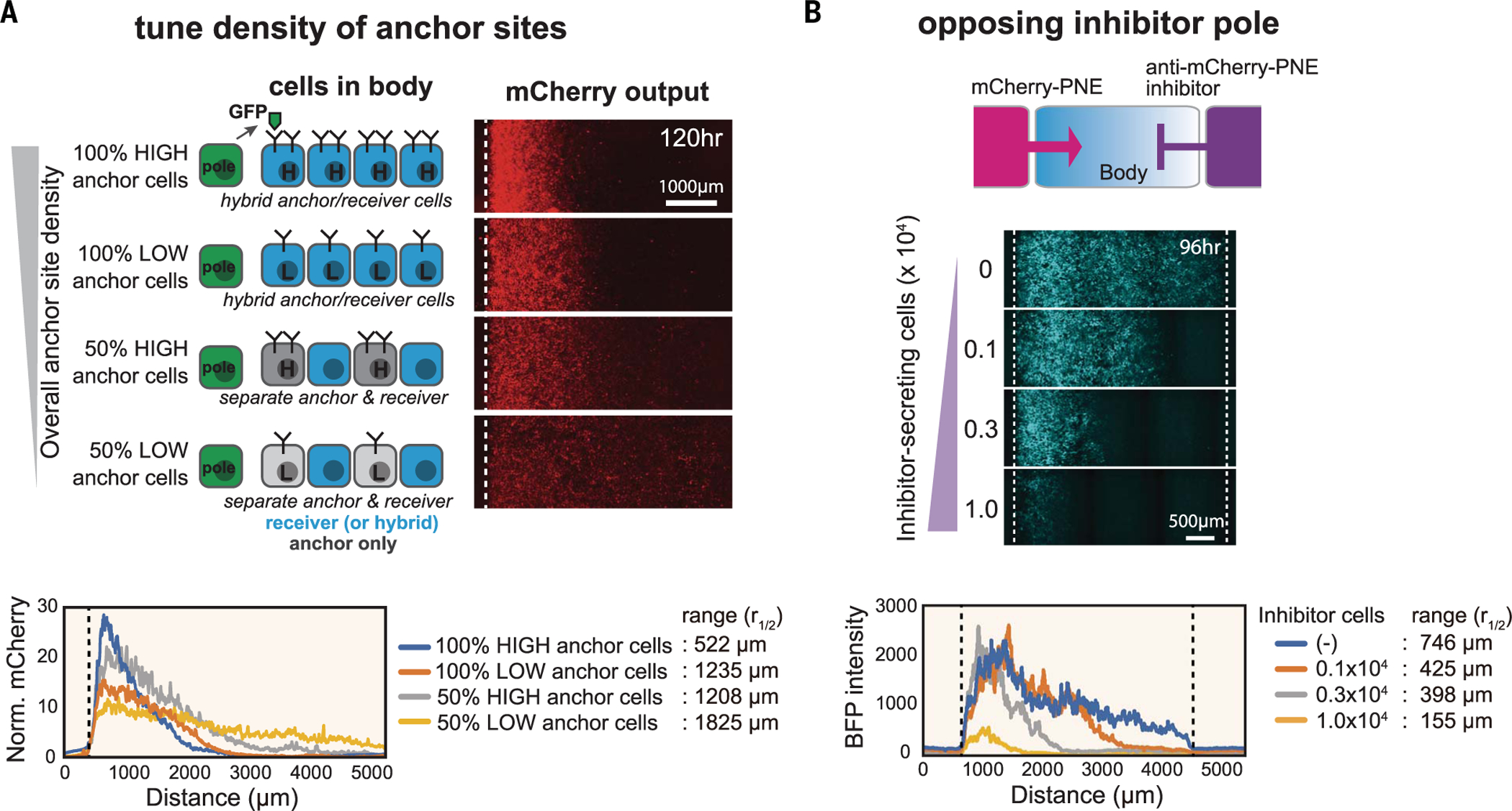
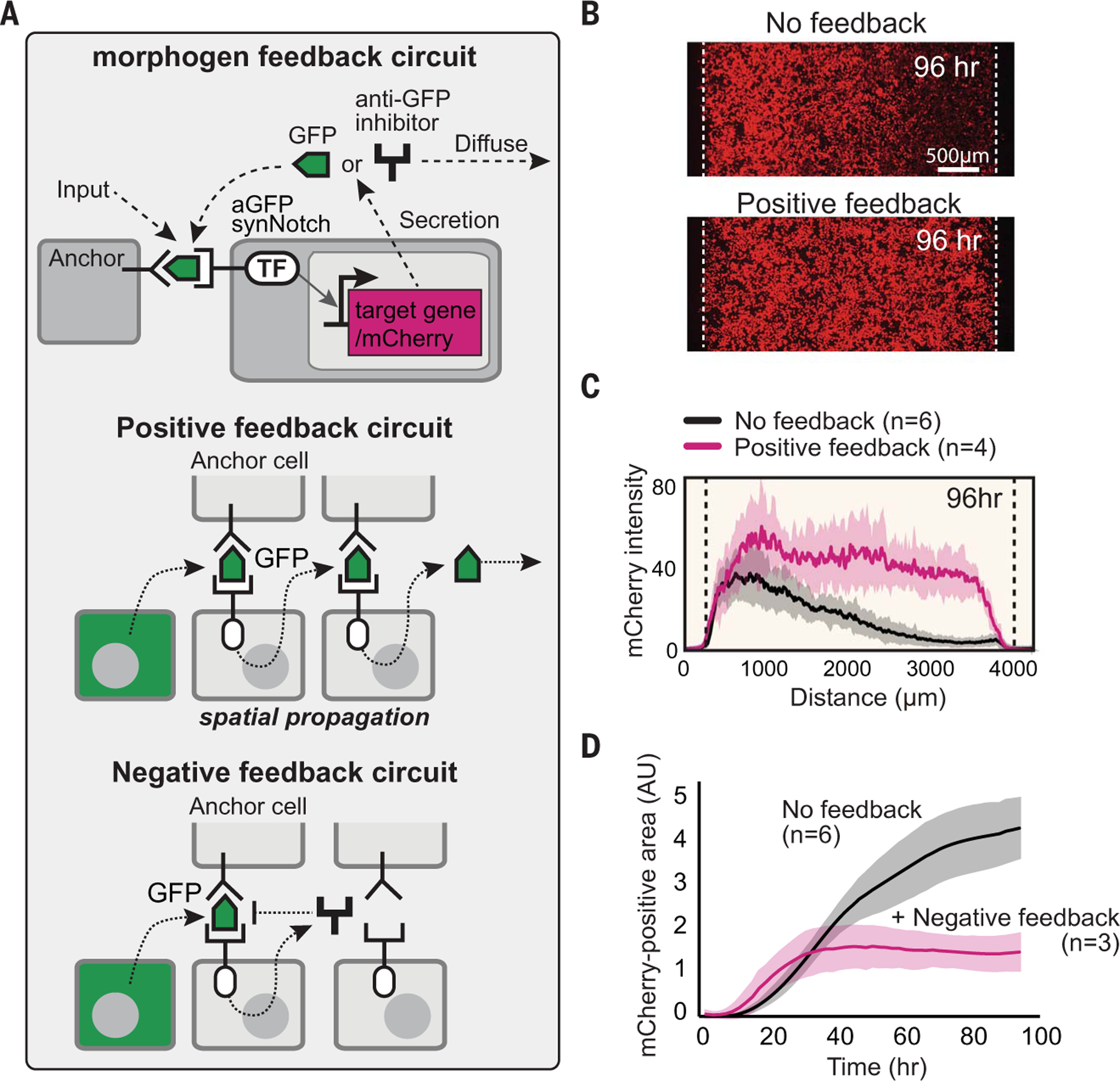
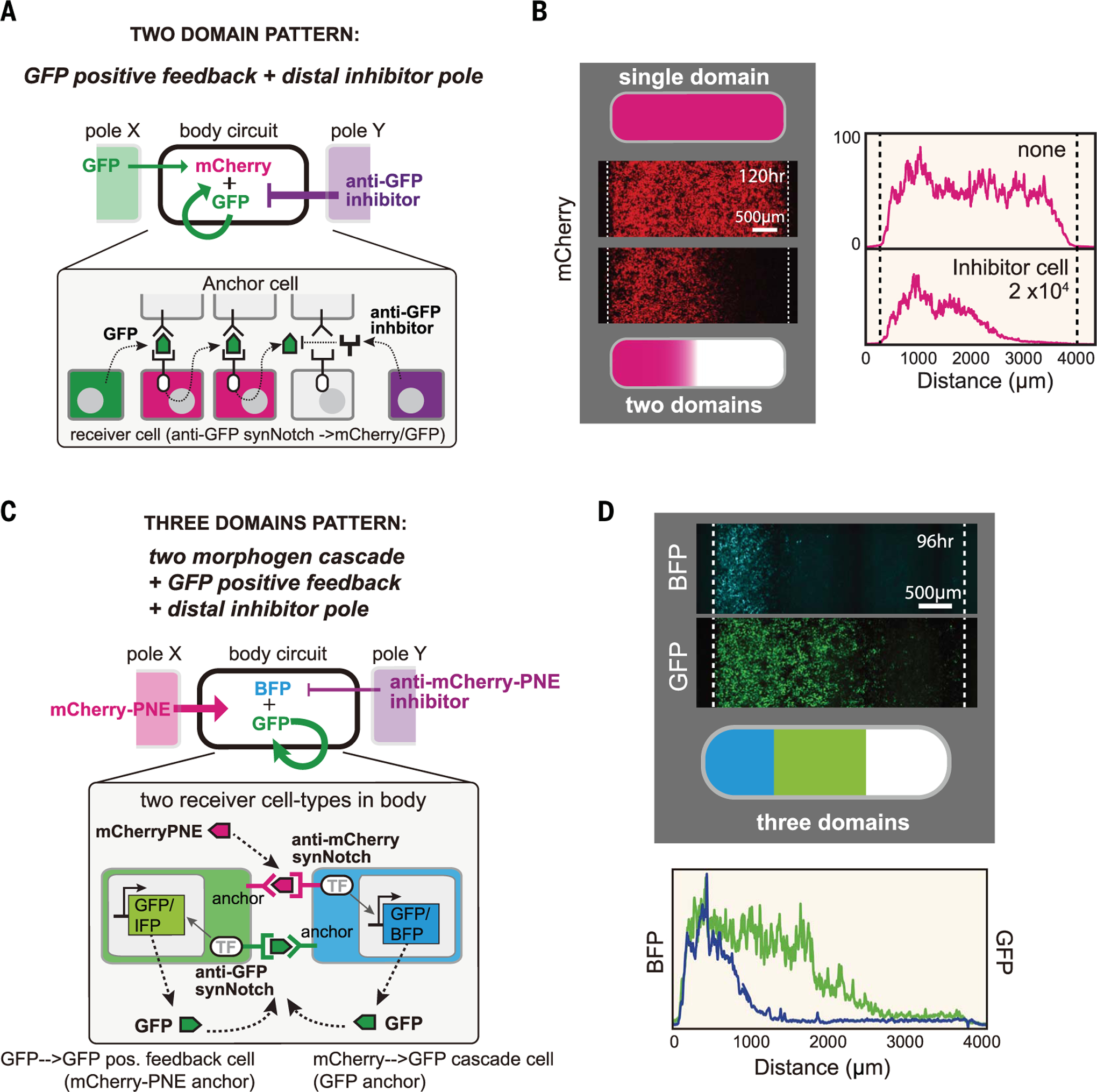
In metazoan tissues, cells decide their fates by sensing positional information provided by specialized morphogen proteins. To explore what features are sufficient for positional encoding, we asked whether arbitrary molecules (e.g., green fluorescent protein or mCherry) could be converted into synthetic morphogens. Synthetic morphogens expressed from a localized source formed a gradient when trapped by surface-anchoring proteins, and they could be sensed by synthetic receptors. Despite their simplicity, these morphogen systems yielded patterns reminiscent of those observed in vivo. Gradients could be reshaped by altering anchor density or by providing a source of competing inhibitor. Gradient interpretation could be altered by adding feedback loops or morphogen cascades to receiver cell response circuits. Orthogonal cell-cell communication systems provide insight into morphogen evolution and a platform for engineering tissues.


Conflict of interest statement

Competing interests: W.A.L. and S.T. have a financial interest in Gilead Biosciences. W.A.L. and S.T. are inventors on a patent application (PCT/US2016/019188) held by the Regents of the University of California that covers binding synthetic Notch receptors.

Figures

Fig. 1.
Fig. 1.. Turning arbitrary proteins into synthetic morphogens.
(A) SynNotch receptors detect juxtacrine signals (e.g., membrane-tethered GFP). In the diffusible synNotch system, soluble GFP is produced from a secretor cell, then trapped by anti-GFP anchor protein, and finally presented to anti-GFP synNotch on a receiver cell. (B) Multiple arbitrary proteins with two recognition sites could be converted into synthetic morphogens (see fig. S3 for construction of mCherry-PNE peptide morphogen). Kd, dissociation constant. (C) Testing diffusible GFP synNotch system in L929 mouse fibroblasts. Anchor cell expresses anti-GFP LaG2 anchor protein. Receiver cell expresses anti-GFP LaG17 synNotch (induces mCherry reporter). 1 × 10⁴ GFP-secreting cells, 0.5 × 10⁴ anchor cells, and 0.5 × 10⁴ receiver cells were cultured overnight, and mCherry induction in receiver cells was measured by flow cytometry. (D) Juxtacrine versus diffusible GFP signaling gradient. Left pole has 3 × 10⁴ sender cells, and right body has 1.5 × 10⁴ cells (100% receiver cells for juxtacrine; 50:50 anchor:receiver cells for diffusible GFP; see fig. S2, A and B). Images were taken by incucyte system over 4 days when system reached steady state (movie S1). Individual lines show mCherry intensity every 12 hours. memb., membrane.
Fig. 2.
Fig. 2.. Systematic control over distance range of synthetic morphogen gradient.
(A) Anchor density can tune synthetic GFP morphogen gradient shape and signaling range. We constructed bodies with four different densities of LaG2-anchor by using two types of variations: anchor expression level (high or low) and fraction of cells in the body that express anchor (100 or 50%). See materials and methods for details. Norm., normalized. (B) Controlling signaling range of mCherry-PNE morphogen with inhibitor. We used a three-well insert wall to build three regions: morphogen pole, body, and inhibitor pole. The morphogen pole has a mixture of 1 × 10⁴ mCherry-PNE–secreting cells and 2 × 10⁴ parental L929 cells. The body has a 1.5 × 10⁴ mixture of anti-PNE anchor cells and anti-mCherry synNotch receiver cells (50:50 ratio). The inhibitor pole has cells expressing anti–mCherry-PNE inhibitor (total cell number: 3 × 10⁴ of cells; varying number of inhibitor cells: 0, 0.1 × 10⁴, 0.3 × 10⁴, or 1.0 × 10⁴; remaining cells were parental L929). BFP output was quantified by In Cell Analyzer 6000 at day 4 (see fig. S6 for GFP inhibitor analysis).
Fig. 3.
Fig. 3.. Reshaping morphogen interpretation with positive or negative feedback.
(A) In a positive feedback circuit, GFP morphogen activates receiver cells to induce the secretion of more GFP. In a negative feedback circuit, GFP morphogen induces the expression of antimorphogen inhibitor by receiver cells. TF, transcription factor. (B) Comparison of mCherry output in the body with and without positive feedback at 96 hours (see fig. S7C for time course). The pole has 3 × 10⁴ GFP-secreting cells; the body has a 1.5 × 10⁴ mixture of anchor cells and receiver cells engineered with a positive feedback circuit (50:50 ratio). Images were taken by incucyte system for 4 days (movie S1). (C) Activity gradient profiles at 96 hours, with and without positive feedback. Shaded area shows SD from multiple experiments. (D) The mCherry-positive area (integral of top plots) plotted over time shows that the body with negative feedback reaches steady state faster than it does without feedback. AU, arbitrary units.
Fig. 4.
Fig. 4.. Combining synthetic morphogen interpretation circuits to engineer multidomain spatial patterns.
(A and B) Programming two-domain pattern by combining positive feedback circuit with opposing morphogen and inhibitor poles. Morphogen pole (X) has 3 × 10⁴ GFP-secreting cells; the body has a 50:50 mixture of anchor cells and receiver cells with positive feedback (used in Fig. 3B) (1.5 × 10⁴ total cells); and the inhibitor pole (Y) has 2 × 10⁴ anti-GFP inhibitor–secreting cells. Images were taken by incucyte at 120 hours (movie S1). See fig. S8B for variant circuits. (C and D) Programming three-domain pattern. We combined the two-morphogen cascade and positive feedback circuit with opposing morphogen and inhibitor poles. The body contains two types of cells: Cell A expresses anti-mCherry synNotch that induces BFP reporter and GFP morphogen (mCherry-PNE→GFP cascade), and cell B expresses anti-GFP LaG17 synNotch that induces expression of GFP morphogen (GFP→GFP positive feedback). See materials and methods for details. Image was taken at 96 hours by In Cell Analyzer 6000 (movie S2). IFP, infrared fluorescent protein.

Comment in

  • Reconstituting tissue patterning.
    Barkai N, Shilo BZ. Barkai N, et al. Science. 2020 Oct 16;370(6514):292-293. doi: 10.1126/science.abe4217. Science. 2020. PMID: 33060349 No abstract available.

References

    1. Turing AM, Phil. Trans. R. Soc. Lond. B 237, 37–72 (1952).
    1. Wolpert L, J. Theor. Biol 25, 1–47 (1969). - PubMed
    1. Rogers KW, Schier AF, Annu. Rev. Cell Dev. Biol 27, 377–407 (2011). - PubMed
    1. Lander AD, Science 339, 923–927 (2013). - PMC - PubMed
    1. Tabata T, Takei Y, Development 131, 703–712 (2004). - PubMed

Publication types

MeSH terms

Substances