. 2020 Oct 15;11(10):863.
doi: 10.1038/s41419-020-03034-3.

Mesenchymal stem cells reverse EMT process through blocking the activation of NF-κB and Hedgehog pathways in LPS-induced acute lung injury

Affiliations

Mesenchymal stem cells reverse EMT process through blocking the activation of NF-κB and Hedgehog pathways in LPS-induced acute lung injury

Kun Xiao et al. Cell Death Dis. .

Abstract

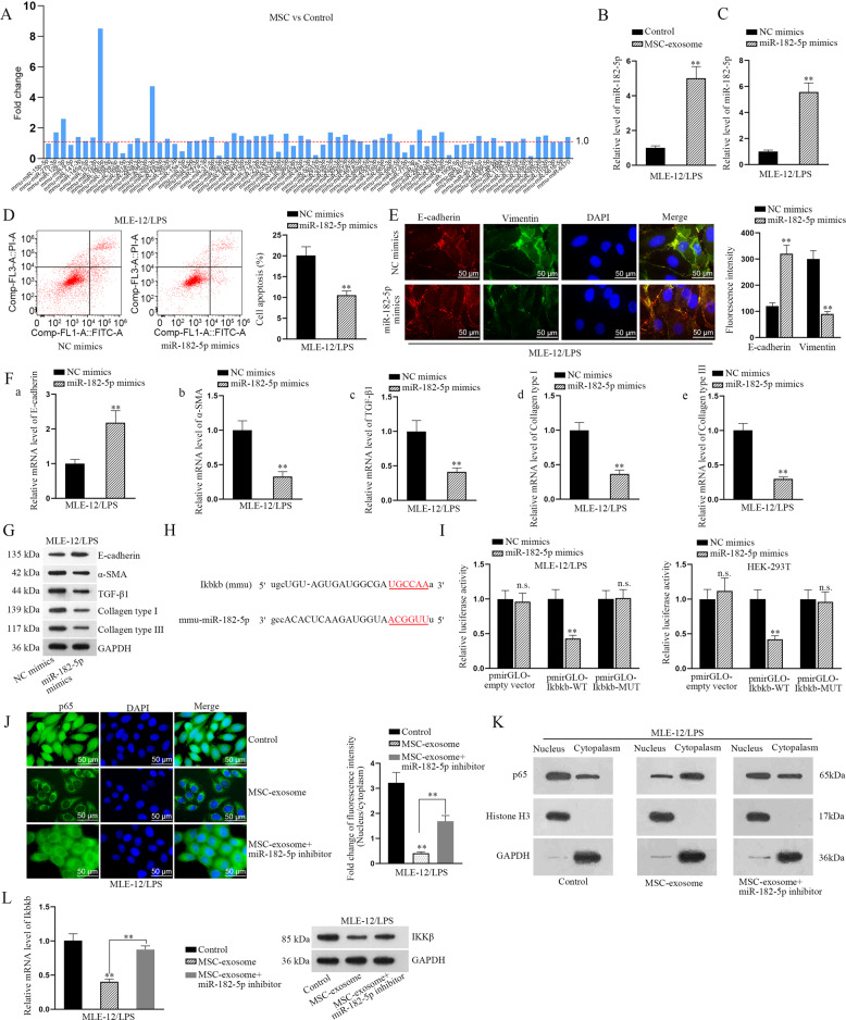
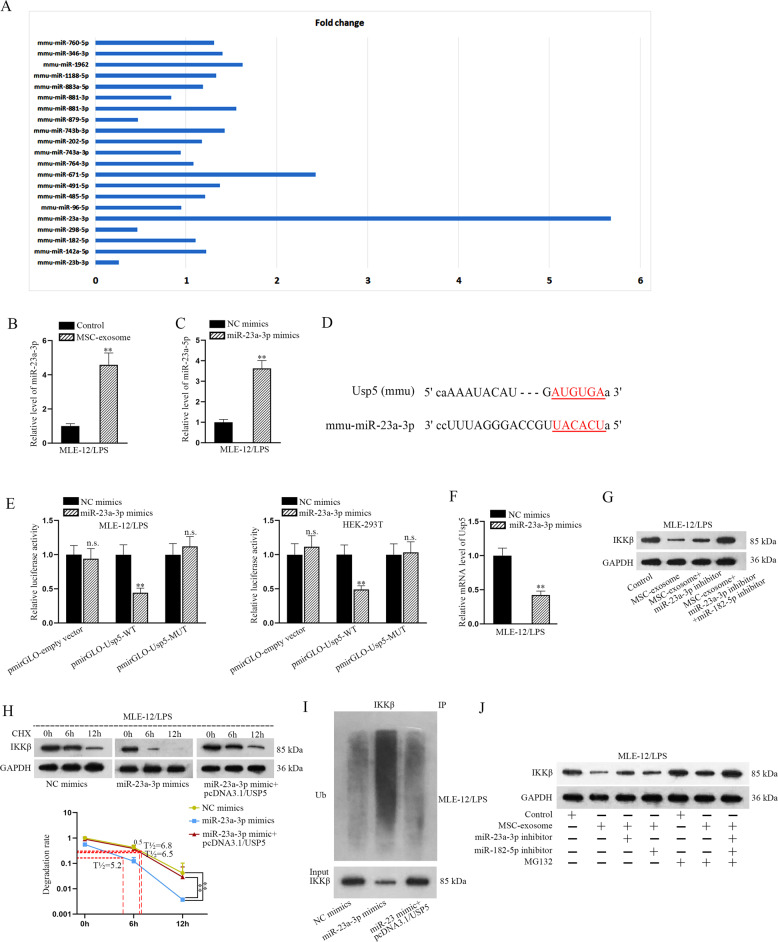
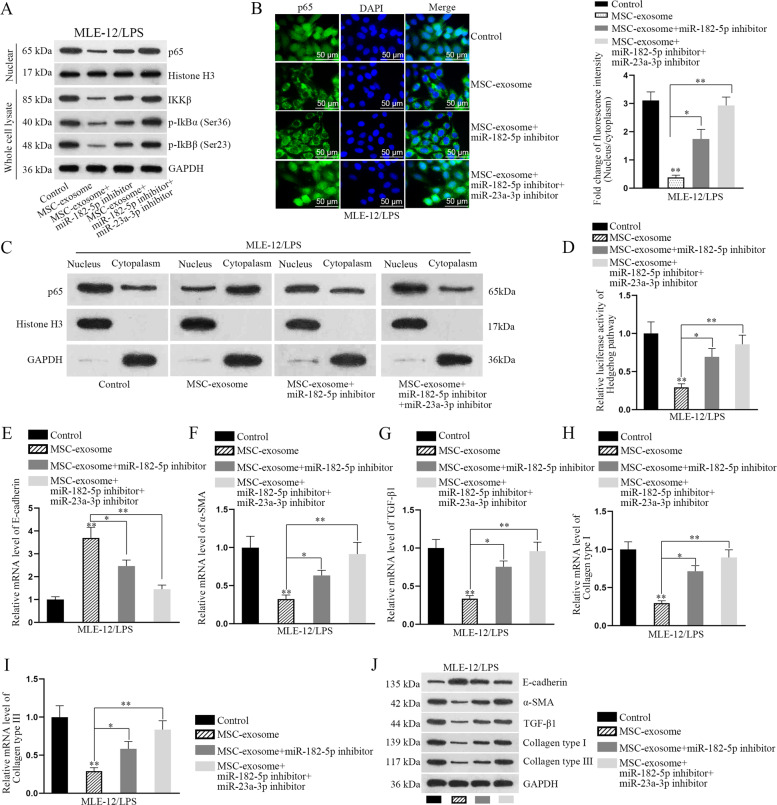
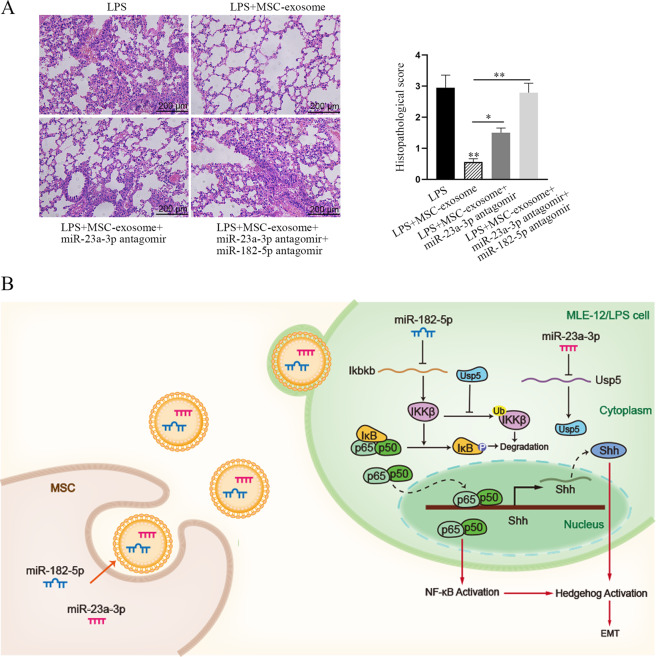
Acute lung injury (ALI) is a pulmonary disorder, which can result in fibrosis of the lung tissues. Recently, mesenchymal stem cell (MSC) has become a novel therapeutic method for ALI. However, the potential mechanism by which MSC regulates the progression of ALI remains blurry. The present study focused on investigating the mechanism underneath MSC-reversed lung injury and fibrosis. At first, we determined that coculture with MSC led to the inactivation of NF-κB signaling and therefore suppressed hedgehog pathway in LPS-treated MLE-12 cells. Besides, we confirmed that MSC-exosomes were responsible for the inhibition of EMT process in LPS-treated MLE-12 cells through transmitting miRNAs. Mechanism investigation revealed that MSC-exosome transmitted miR-182-5p and miR-23a-3p into LPS-treated MLE-12 cells to, respectively, target Ikbkb and Usp5. Of note, Usp5 interacted with IKKβ to hamper IKKβ ubiquitination. Moreover, co-inhibition of miR-182-5p and miR-23a-3p offset the suppression of MSC on EMT process in LPS-treated MLE-12 cells as well as in LPS-injured lungs of mice. Besides, the retarding effect of MSC on p65 nuclear translocation was also counteracted after co-inhibiting miR-182-5p and miR-23a-3p, both in vitro and in vivo. In summary, MSC-exosome transmitted miR-23a-3p and miR-182-5p reversed the progression of LPS-induced lung injury and fibrosis through inhibiting NF-κB and hedgehog pathways via silencing Ikbkb and destabilizing IKKβ.


Conflict of interest statement

The authors declare that they have no conflict of interest.

Figures

Fig. 1
Fig. 1. NF-κB and hedgehog signaling pathways are inactivated in LPS-treated MLE-12 cells cocultured with MSC.
a The cocultured system with LPS-treated MLE-12 cells and MSCs was built. b Luciferase reporter assays detected the relative luciferase activity of various signaling pathways (Notch, Wnt, Nanog, NF-κB, PI3K/AKT, Oct4, Hedgehog, MAPK/JNK, and MAPK/ERK) in LPS-treated MLE-12 cells treated with or without MSC. c The mRNA level of Ikbkb, Chuk, Ikbkg, or RelA was examined by RT-qPCR after cocultured with MSC. d Western blot analysis determined the levels of IKKβ, p-IκBα, and p-IκBβ in the whole cell lysates as well as the nuclear protein level of p65 after cocultured with MSCs. e RT-qPCR analyzed the mRNA levels of hedgehog pathway key factors (Shh, Dhh, Ihh, Ptch1, Smo, and Gli1) in control cells and cocultured cells. f The luciferase activity of hedgehog pathway was detected in cocultured cells. g Relative mRNA level of Shh was measured by RT-qPCR in LPS-treated MLE-12 cells under four different conditions (control, MSC, KINK-1, and KINK-1+MSC). h Relative luciferase activity of hedgehog pathway was measured in LPS-treated MLE-12 cells under the same four conditions. **p < 0.01. n.s. no statistical significance.
Fig. 2
Fig. 2. p65 facilitates the transcriptional activation of Shh in LPS-treated MLE-12 cells.
a RT-qPCR detected the level of Shh in LPS-treated MLE-12 cells with p65 overexpression or inhibition. b ChIP assay examined the enrichment of Shh promoter induced by p65 or IgG in LPS-treated MLE-12 cell. c, d JASPAR predicted the DNA motif of p65 and its binding sites in Shh promoter. e ChIP assay detected the enrichment of Shh promoter on P1 or P2 in p65- or IgG-precipitated compounds in LPS-treated MLE-12 cells. f Luciferase reporter assays detected the luciferase activity of indicated Shh promoter (WT, MUT1, MUT2, and MUT) in LPS-treated MLE-12 cells under p65 overexpression or not. **p < 0.01. n.s. no statistical significance.
Fig. 3
Fig. 3. MSC-secreted exosomes inactivate NF-κB pathway.
a Luciferase reporter assay detected the luciferase activity of Ikbkb promoter vector under coculture of MSC. b, c The relative mRNA level of Dicer and Ikbkb was examined by RT-qPCR in LPS-treated MLE-12 cells cocultured with MSC or Dicer-silenced MSC, respectively. d The protein levels of NF-κB pathway key factors (IKKβ, p-IκBα, and p-IKBβ in the whole cell lysate as well as the nuclear protein level of p65) were checked using western blot after cocultured with MSC or Dicer-silenced MSC. e Electron microscope analyzed the existence of exosomes secreted by MSC or Dicer-silenced MSC. f PKH67 staining was used to confirm the entrance of exosomes secreted by MSC or Dicer-silenced MSC into target cells. Scale bar = 100 μm. g The diameter of exosomes was measured and identified through NTA. h The surface markers of exosomes (CD9, CD63, CD81, and HSP70) were detected by western blot analysis. i RT-qPCR examined the level of Ikbkb mRNA in LPS-treated MLE-12 cells treated with MSC-exosome or MSC/sh-Dicer-exosome. j Western blot examined the protein levels of nuclear p65, Ikkβ, p-IκBα, and p-IκBβ in LPS-treated MLE-12 cells treated with MSC-exosome or MSC/sh-Dicer-exosome. k IF staining detected the nuclear translocation of p65 in LPS-treated MLE-12 cells under the context of MSC-exosome or MSC/sh-Dicer-exosome. Scale bar = 50 μm. l The cellular location of p65 was validated by western blot analysis in LPS-treated MLE-12 cells with MSC-exosome or MSC/sh-Dicer-exosome treatment. **p < 0.01. n.s. no statistical significance.
Fig. 4
Fig. 4. MiR-182-5p transmitted by MSC-exosomes reverses EMT process by directly targeting Ikbkb.
a Online starBase v2.0 predicted 71 miRNAs targeted to Ikbkb were subjected to RT-qPCR analysis in LPS-treated MLE-12 cells under MSC coculture or not. b RT-qPCR analyzed the level of miR-182-5p in LPS-treated MLE-12 cells treated with MSC-exosome. c Relative level of miR-182-5p was detected by RT-qPCR in LPS-treated MLE-12 cells transfected with specific miRNA mimics. d Flow cytometry analysis displayed the apoptosis rate in LPS-treated MLE-12 cells transfected with miR-182-5p mimics. e IF assay analyzed the fluorescence intensities of two EMT-related proteins, E-cadherin, and Vimentin, in LPS-treated MLE-12 cells transfected with miR-182-5p mimics. Scale bar = 50 μm. f, g RT-qPCR and western blot examined the levels of epithelial marker (E-cadherin) and mesenchymal markers (α-SMA, TGF-β1, Collagen type I, and Collagen type III) in LPS-treated MLE-12 cells transfected with miR-182-5p mimics. h The binding sites between miR-182-5p and Ikbkb were predicted. i Luciferase reporter assay examined the luciferase activity of indicated reporter vectors in LPS-treated MLE-12 cells and HEK-293T cells co-transfected with miR-182-5p mimics or NC mimics. j IF assay detected p65 nuclear translocation in LPS-treated MLE-12 cells with MSC-exosome or MSC-exosome+miR-182-5p inhibitor. Scale bar = 50 μm. k Western blot analysis detected cytoplasmic and nuclear p65 in LPS-treated MLE-12 cells with MSC-exosome or MSC-exosome+miR-182-5p inhibitor. l RT-qPCR and western blot examined the mRNA and protein levels of Ikbkb in LPS-treated MLE-12 cells transfected with MSC-exosome or MSC-exosome+miR-182-5p inhibitor. **p < 0.01. n.s. no statistical significance.
Fig. 5
Fig. 5. MSC-exosome induces the ubiquitination of IKKβ through downregulating Usp5.
a After treated with CHX in three different time points, the protein level of IKKβ was tested in LPS-treated MLE-12 cells with or without MSC-exosome. b The protein level of IKKβ was detected in LPS-treated MLE-12 cells treated with control or MSC-exosome or control+MG132 or MSC-exosome+MG132. c Ubiquitination assay detected the ubiquitination of IKKβ protein in LPS-treated MLE-12 cells treated with MSC-exosome. d Pull-down silver staining was applied to unveil the proteins that might interact with IKKβ. e Co-IP assay demonstrated the interaction between Usp5 and IKKβ. f RT-qPCR and western blot examined the overexpression efficiency of Usp5 and the protein level of IKKβ in response to Usp5 overexpression. g Western blot analyzed the protein level of IKKβ in LPS-treated MLE-12 cells under four situations (control, MSC-exosome, MSC-exosome+pcDNA3.1/Usp5, or MSC-exosome+pcDNA3.1/Usp5+miR-182-5p inhibitor). h Luciferase reporter assays indicated the relative luciferase activity of Usp5 promoter in LPS-treated MLE-12 cells treated with MSC-exosome. i RT-qPCR and western blot detected the mRNA level and protein level of Usp5 in LPS-treated MLE-12 cells treated with MSC-exosome or MSC/sh-Dicer-exosome. **p < 0.01. n.s. no statistical significance.
Fig. 6
Fig. 6. MiR-23a-3p transmitted by MSC-exosome regulates the ubiquitination of IKKβ through targeting Usp5.
a StarBase v2.0 predicted miRNAs targeted to Usp5 were subjected to RT-qPCR analysis in LPS-treated MLE-12 cells with or without MSC coculture. b RT-qPCR analyzed miR-23a-3p expression in LPS-treated MLE-12 cells treated with MSC-exosome. c RT-qPCR analyzed miR-23a-3p expression in LPS-treated MLE-12 cells transfected with miR-23a-3p mimics. d The binding sequence between miR-23a-3p and Usp5 was shown. e Luciferase reporter assay examined the luciferase activity of indicated vectors in LPS-treated MLE-12 cells and HEK-293T cells co-transfected with miR-23a-3p mimics or NC mimics. f The expression level of Usp5 was detected by RT-qPCR in LPS-treated MLE-12 cells after the transfection of miR-23a-3p mimics. g Western blot measured the protein level of IKKβ in LPS-treated MLE-12 cells treated with different groups (control, MSC-exosome, MSC-exosome+miR-23a-3p inhibitor or MSC-exosome+miR-23a-3p inhibitor+miR-182-5p inhibitor). h After CHX treatment, the half-life of IKKβ was detected in LPS-treated MLE-12 cells transfected with NC mimics, miR-23a-3p mimics or miR-23a-3p mimics+pcDNA3.1/Usp5. i The ubiquitination level of IKKβ was detected in LPS-treated MLE-12 cells transfected with NC mimics, miR-23a-3p mimics or miR-23a-3p mimics+pcDNA3.1/Usp5. j Western blot examined the protein level of IKKβ in LPS-treated MLE-12 cells under seven conditions (control, MSC-exosome, MSC-exosome+miR-23a-3p inhibitor, MSC-exosome+miR-182-5p inhibitor, control+MG132, MSC-exosome+MG132, and MSC-exosome+miR-23a-3p inhibitor+miR-182-5p inhibitor+MG132). **p < 0.01. n.s. no statistical significance.
Fig. 7
Fig. 7. Exosomal miR-23a-3p and miR-182-5p attenuates LPS-induced injury and EMT process in MLE-12 cells by negatively regulating Usp5/Ikbkb axis.
Rescue assays were carried out in LPS-treated MLE-12 cells under four different contexts (control, MSC-exosome, MSC-exosome+miR-182-5p inhibitor, and MSC-exosome+miR-182-5p inhibitor+miR-23a-3p inhibitor). a The levels of nuclear p65, IKKβ, p-IKBα, and p-IKBβ were detected in LPS-treated MLE-12 cells using western blot. b IF staining examined the nuclear translocation of p65 in LPS-treated MLE-12 cells under diverse conditions. Scale bar = 50 μm. c The protein level of p65 in nucleus or cytoplasm was examined by western blot analysis in LPS-treated MLE-12 cells with MSC-exosome, MSC-exosome+miR-182-5p inhibitor or MSC-exosome+miR-23a-3p inhibitor. d Luciferase reporter assay examined the luciferase activity of hedgehog pathway in LPS-treated MLE-12 cells. ei RT-qPCR detected the mRNA level of E-cadherin, α-SMA, TGF-β1, Collagen type I, and Collagen type III in indicated LPS-treated MLE-12 cells. j Western blot detected the protein level of E-cadherin, α-SMA, TGF-β1, Collagen type I, and Collagen type III in indicated LPS-treated MLE-12 cells. *p < 0.05, **p < 0.01.
Fig. 8
Fig. 8. MSC-exosome delivered miR-182-5p and miR-23a-3p to mitigate LPS-induced ALI and pulmonary fibrosis by targeting Ikbkb/Usp5 signaling.
a HE staining of lung tissues in four different groups (LPS, LPS + MSC-exosome, LPS + MSC-exosome+miR-23a-3p inhibitor, LPS + MSC-exosome+miR-23a-3p inhibitor+miR-182-5p inhibitor). Scale bar = 200 μm. b The concept map demonstrating the role and functional mechanism of MSC on alleviating ALI and pulmonary fibrosis. *p < 0.05, **p < 0.01.

References

    1. Favarin DC, de Oliveira JR, de Oliveira CJ, Rogerio Ade P. Potential effects of medicinal plants and secondary metabolites on acute lung injury. BioMed. Res. Int. 2013;2013:576479. - PMC - PubMed
    1. Butt Y, Kurdowska A, Allen TC. Acute lung injury: a clinical and molecular review. Arch. Pathol. Lab. Med. 2016;140:345–350. doi: 10.5858/arpa.2015-0519-RA. - DOI - PubMed
    1. Blackwell TS, et al. Future directions in idiopathic pulmonary fibrosis research. An NHLBI workshop report. Am. J. Respir. Crit. Care Med. 2014;189:214–222. doi: 10.1164/rccm.201306-1141WS. - DOI - PMC - PubMed
    1. Thannickal VJ, et al. Mechanisms of pulmonary fibrosis. Annu. Rev. Med. 2004;55:395–417. doi: 10.1146/annurev.med.55.091902.103810. - DOI - PubMed
    1. White ES, Lazar MH, Thannickal VJ. Pathogenetic mechanisms in usual interstitial pneumonia/idiopathic pulmonary fibrosis. J. Pathol. 2003;201:343–354. doi: 10.1002/path.1446. - DOI - PMC - PubMed

Publication types