. 2020 Oct 15;11(1):5208.
doi: 10.1038/s41467-020-18952-1.

Topaz-Denoise: general deep denoising models for cryoEM and cryoET

Affiliations

Topaz-Denoise: general deep denoising models for cryoEM and cryoET

Tristan Bepler et al. Nat Commun. .

Abstract

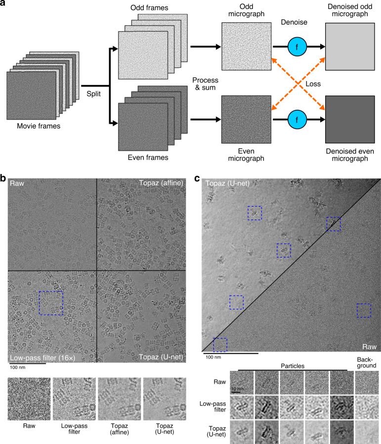
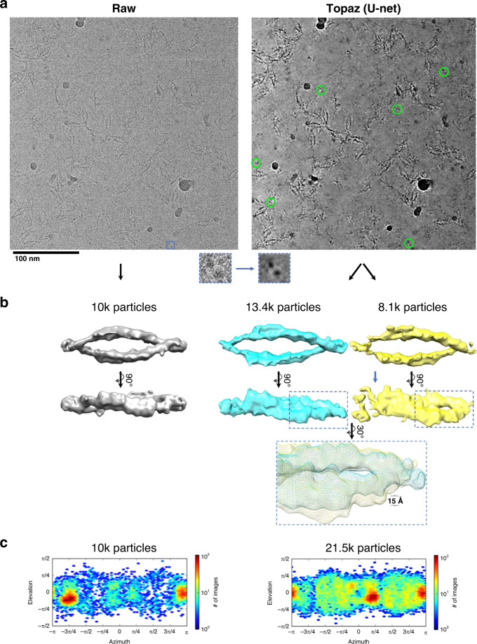
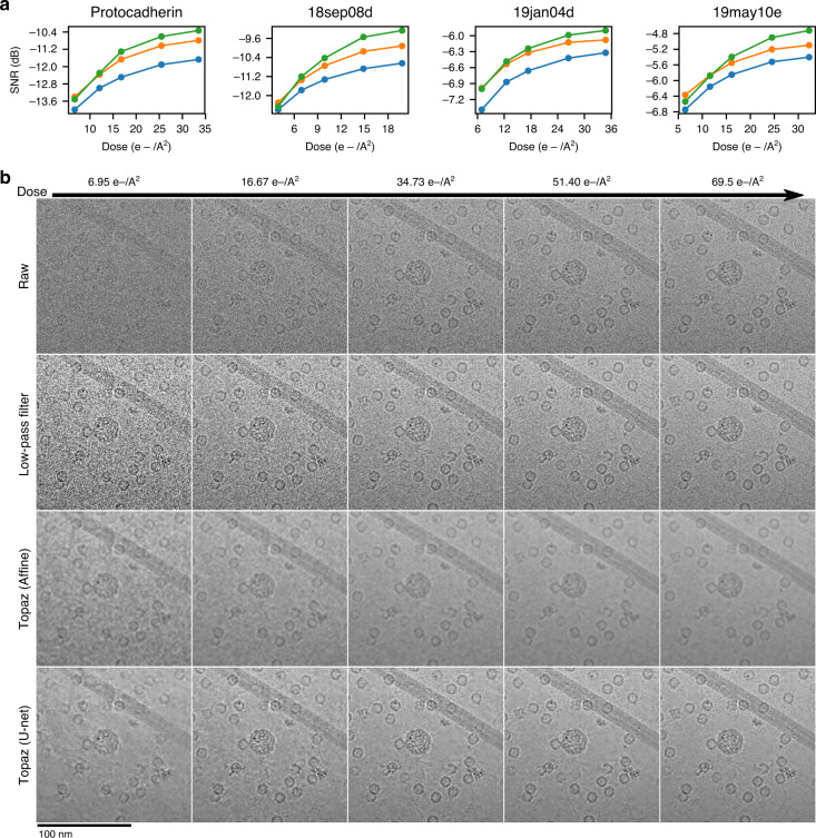
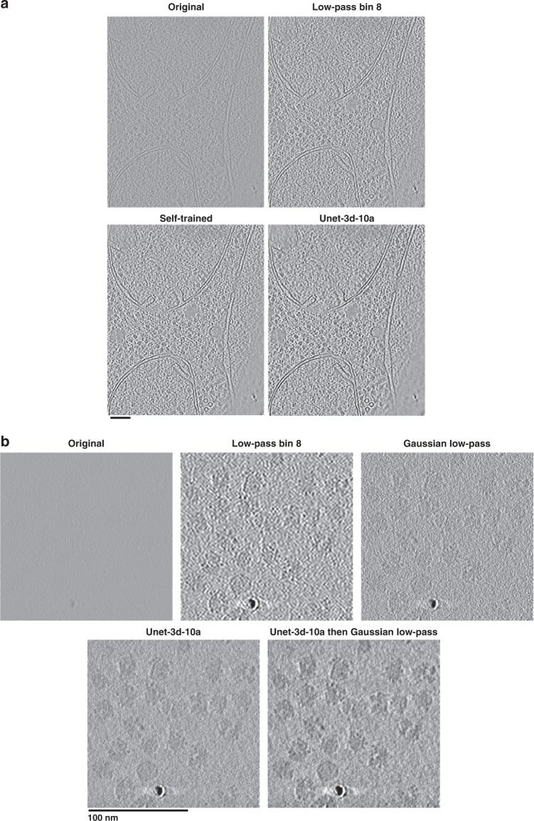
Cryo-electron microscopy (cryoEM) is becoming the preferred method for resolving protein structures. Low signal-to-noise ratio (SNR) in cryoEM images reduces the confidence and throughput of structure determination during several steps of data processing, resulting in impediments such as missing particle orientations. Denoising cryoEM images can not only improve downstream analysis but also accelerate the time-consuming data collection process by allowing lower electron dose micrographs to be used for analysis. Here, we present Topaz-Denoise, a deep learning method for reliably and rapidly increasing the SNR of cryoEM images and cryoET tomograms. By training on a dataset composed of thousands of micrographs collected across a wide range of imaging conditions, we are able to learn models capturing the complexity of the cryoEM image formation process. The general model we present is able to denoise new datasets without additional training. Denoising with this model improves micrograph interpretability and allows us to solve 3D single particle structures of clustered protocadherin, an elongated particle with previously elusive views. We then show that low dose collection, enabled by Topaz-Denoise, improves downstream analysis in addition to reducing data collection time. We also present a general 3D denoising model for cryoET. Topaz-Denoise and pre-trained general models are now included in Topaz. We expect that Topaz-Denoise will be of broad utility to the cryoEM community for improving micrograph and tomogram interpretability and accelerating analysis.


Conflict of interest statement

The authors declare no competing interests.

Figures

Fig. 1
Fig. 1. Illustration of the training framework and comparison of denoising methods on two example micrographs.
a The Noise2Noise method requires paired noisy observations of the same underlying signal. We generate these pairs from movie frames collected in the normal cryoEM process, because each movie frame is an independent sample of the same signal. These are first split into even/odd movie frames. Then, each is processed and summed independently following standard micrograph processing protocols. The resulting even and odd micrographs are denoised with the denoising model (denoted here as f). Finally, to calculate the loss, the odd denoised micrograph is compared with the raw even micrograph and vice versa. b Micrograph from EMPIAR-10025 split into four quadrants showing the raw micrographs, low-pass filtered micrograph by a binning factor of 16, and results of denoising with our affine and U-net models. Particles become clearly visible in the low-pass filtered and denoised micrographs, but the U-net denoising shows strong additional smoothing of background noise. A detail view of the micrograph is highlighted in blue and helps to illustrate the improved background smoothing provided by our U-net denoising model. c Micrograph from EMPIAR-10261 split into the U-net denoised and raw micrographs along the diagonal. Detailed views of five particles and one background patch are boxed in blue. The Topaz U-net reveals particles and reduces background noise.
Fig. 2
Fig. 2. Denoising with the general model in Topaz improves interpretability and picking of difficult particle projections.
a A raw micrograph (left) and Topaz-Denoised micrograph (right) of the clustered protocadherin dataset (EMPIAR-10234) with a top-view boxed out (insets). Denoising allows for top-views to be clearly identified (green circles, right) and subsequently used to increase the confidence and completion of particle picking. b Topaz picking training on raw micrographs using 1540 manually picked particles from the raw micrographs resulted in the reconstruction on the left. Topaz picking training on the raw micrographs using 1023 manually picked particles from the denoised micrographs resulted in the reconstruction on the right. Manually picking on denoised micrographs resulted in 115% more particles in the 3D reconstruction, which allowed for classification into a closed (blue) and putative partially open (yellow; blue arrow showing disjoint) conformation. The inset shows a zoom-in of the ~15 Å conformational change of the twist. c 3D reconstruction particle distributions for (left) Topaz picking training on raw micrographs using 1540 manually picked particles from the raw micrographs, and (right) Topaz picking training on the raw micrographs using 1,023 manually picked particles from the denoised micrographs. All particles from the two classes in (b, right) are shown (c, right). 3DFSC plots for the three maps shown here are in Supplementary Fig. 14.
Fig. 3
Fig. 3. Denoising with the general model in Topaz enhances SNR of short exposure micrographs.
a SNR (dB) calculated using the split-frames method (see Methods) as a function of electron dose in low-pass filtered micrographs by a binning factor of 16 (blue), affine denoised micrographs (orange), and U-net denoised micrographs (green) in the four NYSBC K2 datasets. Our U-net denoising model enhances the SNR of micrographs across almost all dosages in all four datasets. U-net denoising enhances SNR by a factor of 1.5× or more over low-pass filtering at 20 e-/A2. b Example section of a micrograph from the 19jan04d dataset of apoferritin, β-galactosidase, a VLP, and TMV (full micrograph in Supplementary Figs. 3 and 4) showing the raw micrograph, low-pass filtered micrograph, affine denoised micrograph, and U-net denoised micrograph over increasing dose. Particles are clearly visible at the lowest dose in the denoised micrograph and background noise is substantially reduced by Topaz denoising.
Fig. 4
Fig. 4. Denoising with the 3D general model in Topaz improves cellular and single particle tomogram interpretability.
a Saccharomyces uvarum lamellae cryoET slice-through (collected at 6 Å pixelsize and 18 microns defocus, then binned by 2). The general denoising model (Unet-3d-10a) is comparable visually and by SNR to the model trained on the tomogram’s even/odd halves (Self-trained). Both denoising models show an improvement in protein and membrane contrast over binning by 8 while confidently retaining features of interest, such as proteins between membrane bilayers. Both denoising models also properly smooth areas with minimal protein/membrane density compared to the binning by 8. See Supplementary Movie 1 for the tomogram slice-throughs. b 80S ribosomes as single particles (EMPIAR-10045; collected at 2.17 Å pixelsize and 4 microns defocus). The general denoising model (Unet-3d-10a) is markedly improved over binning by 8 and the 1/8 Nyquist Gaussian low-pass, both with smoothing background appropriately while increasing contrast and with retaining features of interest at high fidelity, such as the RNA binding pocket in all orientations. The same 1/8 Nyquist Gaussian low-pass applied to the denoised tomogram further improves contrast by suppressing high-frequencies that the user may deem unimportant. See Supplementary Movie 2 for the tomogram slice-throughs.

References

    1. Baxter WT, Grassucci RA, Gao H, Frank J. Determination of signal-to-noise ratios and spectral SNRs in cryo-EM low-dose imaging of molecules. J. Struct. Biol. 2009;166:126–132. doi: 10.1016/j.jsb.2009.02.012. - DOI - PMC - PubMed
    1. Sindelar CV, Grigorieff N. An adaptation of the Wiener filter suitable for analyzing images of isolated single particles. J. Struct. Biol. 2011;176:60–74. doi: 10.1016/j.jsb.2011.06.010. - DOI - PMC - PubMed
    1. Penczek, P. A. Chapter Two—Image Restoration in Cryo-Electron Microscopy. In Methods in Enzymology (ed. Jensen, G. J.) Vol. 482, 35–72 (Academic Press, 2010). - PMC - PubMed
    1. Merk A, et al. Breaking Cryo-EM resolution barriers to facilitate drug discovery. Cell. 2016;165:1698–1707. doi: 10.1016/j.cell.2016.05.040. - DOI - PMC - PubMed
    1. Herzik MA, Wu M, Lander GC. High-resolution structure determination of sub-100 kDa complexes using conventional cryo-EM. Nat. Commun. 2019;10:1–9. doi: 10.1038/s41467-019-08991-8. - DOI - PMC - PubMed

Publication types