. 2020 Oct 15;10(1):17446.
doi: 10.1038/s41598-020-74296-2.

Explosive-driven double-blast exposure: molecular, histopathological, and behavioral consequences

Affiliations

Explosive-driven double-blast exposure: molecular, histopathological, and behavioral consequences

Erin K Murphy et al. Sci Rep. .

Abstract

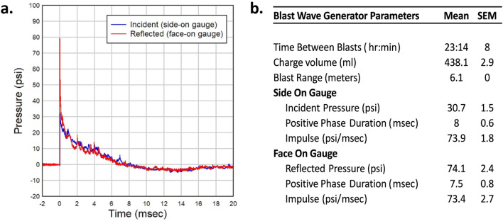
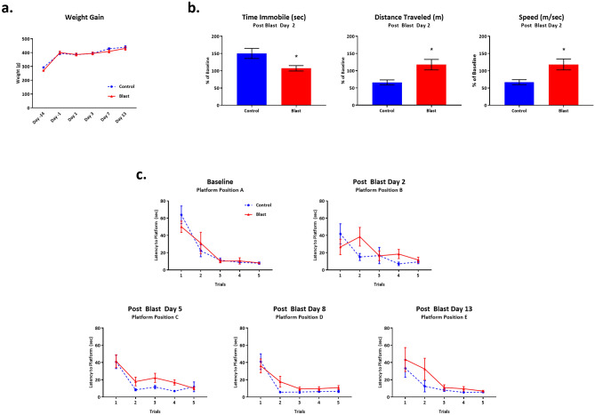
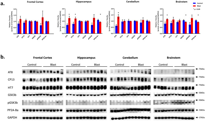
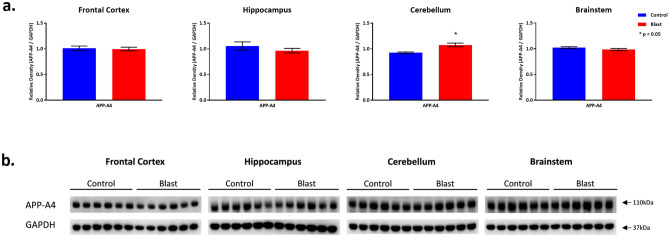
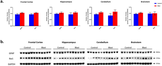
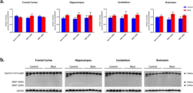
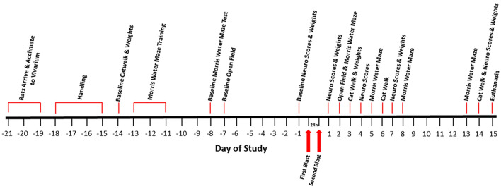
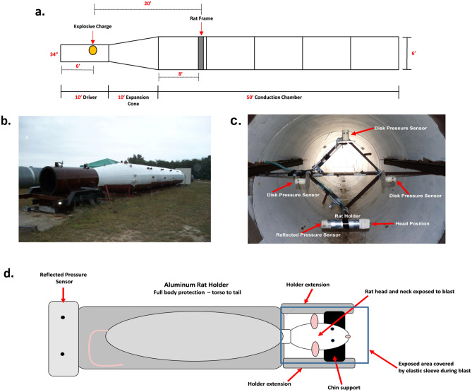
Traumatic brain injury generated by blast may induce long-term neurological and psychiatric sequelae. We aimed to identify molecular, histopathological, and behavioral changes in rats 2 weeks after explosive-driven double-blast exposure. Rats received two 30-psi (~ 207-kPa) blasts 24 h apart or were handled identically without blast. All rats were behaviorally assessed over 2 weeks. At Day 15, rats were euthanized, and brains removed. Brains were dissected into frontal cortex, hippocampus, cerebellum, and brainstem. Western blotting was performed to measure levels of total-Tau, phosphorylated-Tau (pTau), amyloid precursor protein (APP), GFAP, Iba1, αII-spectrin, and spectrin breakdown products (SBDP). Kinases and phosphatases, correlated with tau phosphorylation were also measured. Immunohistochemistry for pTau, APP, GFAP, and Iba1 was performed. pTau protein level was greater in the hippocampus, cerebellum, and brainstem and APP protein level was greater in cerebellum of blast vs control rats (p < 0.05). GFAP, Iba1, αII-spectrin, and SBDP remained unchanged. No immunohistochemical or neurobehavioral changes were observed. The dissociation between increased pTau and APP in different regions in the absence of neurobehavioral changes 2 weeks after double blast exposure is a relevant finding, consistent with human data showing that battlefield blasts might be associated with molecular changes before signs of neurological and psychiatric disorders manifest.


Conflict of interest statement

The authors declare no competing interests.

Figures

Figure 1
Figure 1
Blast Wave characteristics. (a) A representative pressure vs time waveform as produced by detonation of explosives within the Blast Wave Generator and measured with both side-on and face-on recording gauges. This waveform demonstrates a peak incident overpressure around 30 psi with a positive phase duration of approximately 8 ms. (b) Summary output data from Blast Wave Generator as recorded at time of blast.
Figure 2
Figure 2
Behavioral outcomes following Double Blast Exposure. (a) No significant differences in weight gain were noted between 2 × B and Ctl over the course of the study, n = 12 per group. (b) Two days following the second exposure to blast, Open Field analysis revealed that blast exposed rats traveled significantly further, with greater speed, and spent less time immobile during the 5 min test session as compared with controls, n = 12 per group. * indicates p value < 0.05 as determined by 2-tailed, unpaired t test. Bars represent % of baseline for each group. Error bars represent standard error of the mean. (c) No significant differences in latency to find the hidden platform on the Morris water maze were noted between 2 × B and Ctl rats at all time points. n = 12 per group.
Figure 3
Figure 3
Tau and Related protein expression changes following Double Blast Exposure. (a) Histograms representing the densitometric ratio of levels of pTau (phosphorylated-Tau; AT8, CP13), total-Tau (HT7), kinases (GSK3β and pGSK3β), and phosphatase (PP2A-Bα) with respect to GAPDH as measured in the frontal cortex, hippocampus, cerebellum and brainstem in the brains of rats 15-days after an explosive-driven double blast (24 h apart) exposure, n = 6 per group, * indicates p values < 0.05 as determined by 2-tailed, unpaired, t-tests. Error bars represent standard error of the mean (SEM). (b) Representative western blots* for each antibody used. *For full length blots for each antibody, see Supplementary Fig. S10–S13 online.
Figure 4
Figure 4
APP-A4 protein expression following Double Blast Exposure. (a) Histograms representing the densitometric ratio of levels of APP-A4 with respect to GAPDH as measured in the frontal cortex, hippocampus, cerebellum and brainstem in the brains of rats 15-days after an explosive-driven double blast (24 h apart) exposure, n = 6 per group. * indicates p values < 0.05 as determined by 2-tailed, unpaired, t-tests. Error bars represent standard error of the mean (SEM). (b) Representative western blots* for antibodies used. *For full length blots for each antibody, see Supplementary Fig. S10–S13 online.
Figure 5
Figure 5
GFAP and Iba1 protein expression following Double Blast Exposure. (a) Histograms representing the densitometric ratio of levels of GFAP and Iba1 with respect to GAPDH as measured in the frontal cortex, hippocampus, cerebellum and brainstem in the brains of rats 15-days after an explosive-driven double blast (24 h apart) exposure, n = 6 per group. Error bars represent standard error of the mean (SEM). (b) Representative western blots* for each antibody used. *For full length blots for each antibody, see Supplementary Fig. S10–S13 online.
Figure 6
Figure 6
Spectrin and spectrin breakdown product expression following Double Blast Exposure. (a) Histograms representing the densitometric ratio of levels of full length spectrin (240kD) and spectrin breakdown products (SBDP 150kD and 120kD) with respect to GAPDH as measured in the frontal cortex, hippocampus, cerebellum and brainstem in the brains of rats 15-days after an explosive-driven double blast (24 h apart) exposure, n = 5* to 6 per group. Error bars represent standard error of the mean (SEM). *Only 5 control samples available in this experiment. (b) Representative western blots* for each antibody used. *For full length blots for each antibody, see Supplementary Fig. S10–S13 online.
Figure 7
Figure 7
Experimental Timeline. A schematic diagram of the time course for this study. Time interval between blasts is 24 h.
Figure 8
Figure 8
Blast tube parameters. (a) A diagrammatic scheme of the blast-tube used for this study. (b) The exterior aspect of the explosive-driven blast-tube used for this experiment. (c) A cross section of the inside of the BWG at the level of the rat holder and pressure gauges. This picture is taken from the perspective of the explosive charge located in the driver section of the BWG. In this picture, the rat holder is placed on the lower corner of the frame; disk pressure gauges are placed on the remaining three corners for the recording of incident (side-on) pressure. A “face-on” gauge, for the recording of reflected pressure, is fitted at the tail end of the rat holder (visible as a small spot at the left end of the holder in the picture). Rat holder and pressure gauges are equidistant from the explosive charge. During the actual blast exposures, rats were placed 2 at a time in the frame at the top and bottom vertices with the disk pressure gauges on the right and left vertices. (d) Schematic of the aluminum rat holder and rat position within the holder from the perspective of the explosive charge.

References

    1. Elder G, Mitsis E, Ahlers S, Cristian A. Blast-induced mild traumatic brain injury. Psychiatr. Clin. North Am. 2010;33:757–781. doi: 10.1016/j.psc.2010.08.001. - DOI - PubMed
    1. Hoge C, et al. Mild traumatic brain injury in U.S. soldiers returning from Iraq. N. Engl. J. Med. 2008;358:453–463. doi: 10.1056/NEJMoa072972. - DOI - PubMed
    1. Wallace D. Improvised explosive devices and traumatic brain injury: the military experience in Iraq and Afghanistan. Australas. Psychiatry. 2009;17:218–224. doi: 10.1080/10398560902878679. - DOI - PubMed
    1. Rosenfeld J, et al. Blast-related traumatic brain injury. Lancet Neurol. 2013;12:882–893. doi: 10.1016/S1474-4422(13)70161-3. - DOI - PubMed
    1. Abbotts R, Harrison S, Cooper G. Primary blast injuries to the eye: a review of the evidence. J. R. Army Med. Corps. 2007;153:119–123. doi: 10.1136/jramc-153-02-10. - DOI - PubMed

Publication types